
Eye diseases are relatively common in rabbits.
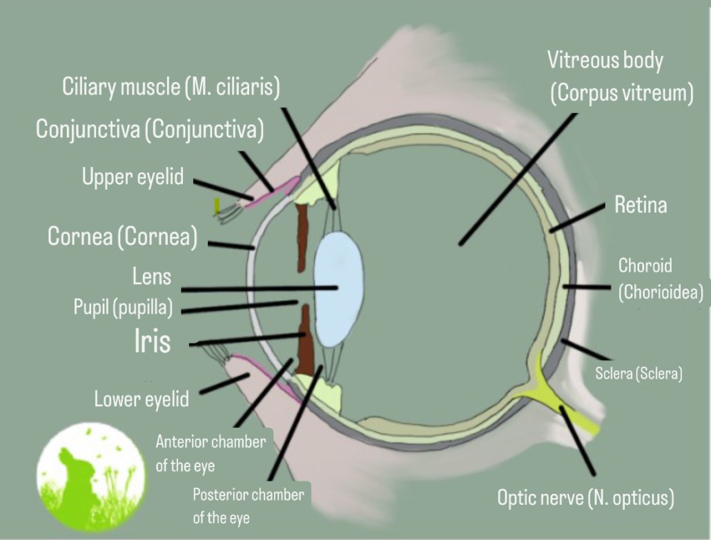
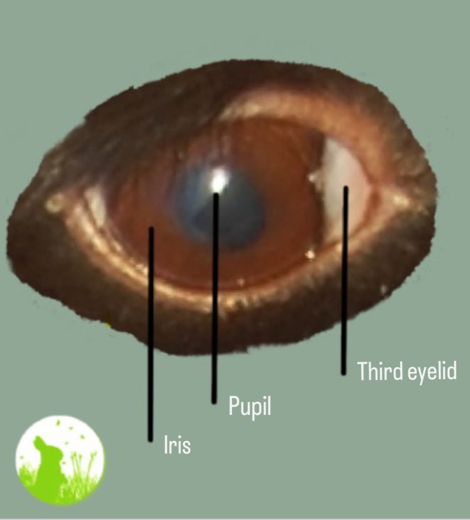
Many eye conditions are treated in ways similar to those in humans or other animals, but some diseases are specific to rabbits, such as inflammation of the choroid associated with E. cuniculi. Understanding the basic structure of the eye can help explain which part is affected and how certain eye diseases develop.
One of the challenges with eye disease in rabbits is that the severity is often underestimated. Anyone who has experienced eye inflammation or another painful eye condition knows how uncomfortable it can be. Rabbits, however, often continue to behave relatively normally despite significant pain, which can make their discomfort difficult to recognize. Adequate pain management is therefore an important part of treatment.
Some symptoms, such as eye discharge, may also be mistaken for something harmless even though they can require prompt veterinary attention. Delayed treatment may allow the condition to become chronic.
Early diagnosis and treatment by a veterinarian experienced in rabbit medicine are therefore important
We recommend consulting an eye specialist (veterinary ophthalmologist) for eye diseases.
For eye discharge or a protruding/bulging eye, a dental veterinarian specializing in rabbits is more suitable. In the case of uveitis, it is particularly important to ensure the veterinarian is knowledgeable about rabbits.
Care
Creating a suitable environment can support the healing of eye conditions. Avoid drafts or wind, direct sunlight or bright light, as well as smoky, dry or cold air, as these conditions are not suitable during recovery.
Preliminary Examination
You can inspect the eye before visiting the veterinarian. Check whether any injuries are visible, whether the problem affects the eye itself or only the eyelid margin, and whether any foreign objects, such as strands of hay, can be seen. To look behind the lower eyelid, gently pull it downward using light pressure.
However, foreign objects should not be removed by non-professionals. Using tweezers can easily injure the eye, especially because rabbits rarely remain completely still. A veterinarian who is experienced with such procedures can remove the foreign object safely.
Caution: A first visual impression is not enough for an accurate assessment. Many corneal injuries are not visible to the naked eye, and foreign objects can easily be missed.
Administering Medications
Your veterinarian will explain how to administer the prescribed medication. Eye drops and ointments are commonly used. Drops are often more comfortable for the rabbit and easier to apply, while ointments remain in the eye longer and therefore usually need to be applied less frequently.
The least stressful method is usually to administer the medication while the rabbit remains on the floor. Kneel down and position the rabbit gently between your thighs to keep it steady. Avoid lifting the rabbit, as this may cause unnecessary stress. Apply the medication as quickly and gently as possible.
Eye Flushing
To flush the eye, you can use artificial tears, lukewarm boiled water or saline solution. Use a cotton pad soaked in the solution and wipe from the outer corner of the eye toward the inner corner.
Use each cotton pad for only one wipe and replace it before wiping again to avoid contamination.
When to Visit the Veterinarian?
Eye diseases in rabbits are often extremely painful, and delaying treatment can worsen the condition and, in some cases, lead to permanent damage to the eye. For this reason, it is generally advisable to consult an emergency veterinarian promptly for any eye-related problem.
Possible exceptions may include mild eye discharge without blinking, squinting, excessive closing of the eye, or changes in behavior, as well as known recurring eye conditions.
Important: If treatment is provided by an emergency veterinarian who is not experienced with rabbits, make sure that prescribed eye medications do not contain cortisone, such as dexamethasone or prednisolone, unless cortisone is being used specifically to treat a diagnosed uveitis.
Blind Rabbits?
It is not uncommon for rabbits to become blind in one or both eyes. Unlike humans, rabbits do not rely primarily on vision and depend heavily on their other senses.
Rabbits that are blind in one eye have a reduced field of vision but otherwise often behave normally in everyday life.
Even rabbits that are blind in both eyes can adapt surprisingly well. However, it is important to keep their environment as familiar as possible. Avoid moving them unnecessarily, placing sharp or dangerous objects in their surroundings, or frequently rearranging their living space, as these changes can cause stress or confusion.
Important: If a rabbit becomes blind, it should be examined by a veterinarian. The underlying cause may be painful, for example increased intraocular pressure (glaucoma), and may require prompt treatment.
Pigment Flecks
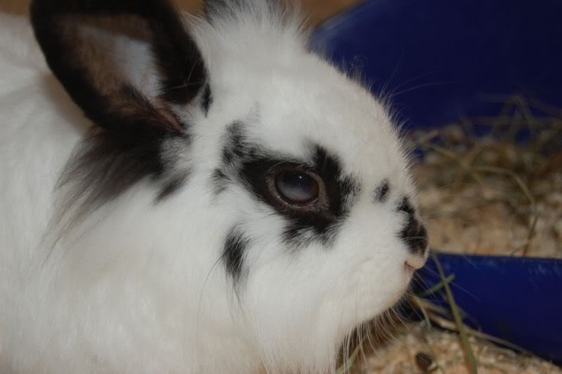
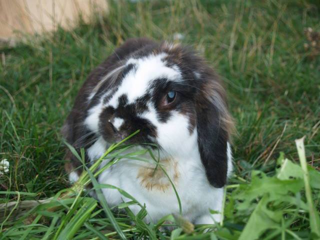
Some rabbits have differently colored pigment spots in their eyes (heterochromia). These may be congenital or genetically determined, but they can also be acquired, for example as a result of delayed or untreated keratitis (corneal inflammation) or eye injuries.
Affected rabbits may show a speckled or mottled appearance in or around the eye.
It is important to distinguish these pigment changes from other eye conditions, such as acute keratitis.
Inflammation or Blockage of the Nasolacrimal Duct (Dacryocystitis)
Eye Discharge
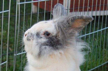
Dacryocystitis is an inflammation or blockage of the tear drainage system, including the tear ducts, nasolacrimal duct, and/or tear sac. It can cause watery or whitish-milky discharge from the eye, often leading to crusting of the surrounding fur and a persistently damp eye area.
If the discharge is clear, this usually indicates a non-infectious cause. In more advanced cases, thick white pus may be present. This pus can also originate from abscesses around the tooth roots that extend into the nasolacrimal duct. Other structures of the eye may also become inflamed, including the cornea, eyelids, and conjunctiva.
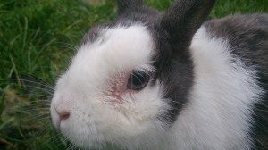
The causes of nasolacrimal duct discharge or inflammation are most commonly dental problems, especially involving the first two molars or the incisors, or bacterial infections. In rabbits, the tooth roots lie very close to the nasolacrimal duct. If the teeth become too long due to insufficient wear or retrograde growth, or if they are misaligned, the tooth roots may enlarge or swell and obstruct the duct.
In many cases, inflamed tooth roots can lead to infection and pus within the nasolacrimal duct, or the infection may spread in the opposite direction.
More information: Jaw Abscesses
Anatomy of the Tear Duct and Its Relationship to Dental Health
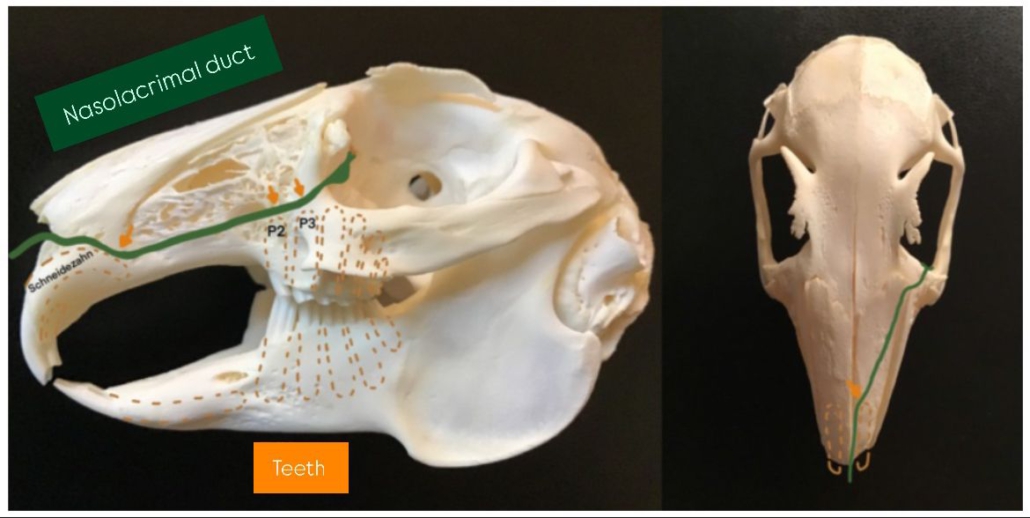
The line shows the course of the tear duct from the tear opening at the eye to the nose. The brown area indicates the part of the teeth that is not visible. The tear duct runs very close to the tooth roots (marked by the orange arrows). For this reason, diseases of the upper jaw, such as retrograde tooth growth or jaw abscesses, often become noticeable only through nasal or eye discharge.
Rabbits with very round heads, such as dwarf rabbits and lop breeds, often have hereditary dental problems that can affect the tear duct. This is related to brachycephaly (a shortened skull shape).
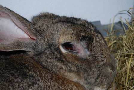
Eye discharge may not become noticeable until a rabbit’s companion passes away. Owners sometimes describe this as the rabbit “crying.” This can happen because the companion rabbit previously cleaned away the discharge several times a day. After the companion dies, the discharge becomes visible. In some cases, the discharge may seem to disappear after the rabbit is paired with a new companion because the new partner cleans it away, making it less noticeable.
The veterinarian will usually first attempt to flush the tear duct, although the duct is not always passable. If it is blocked, dental disease is typically the cause. The tooth roots may grow into the tear duct or compress it due to swelling or inflammation. If the condition remains untreated for a long time, chronic inflammation may cause the tear duct to become blocked or widened, or secretions may clog it.
The cause of the obstruction can be determined more accurately by introducing contrast agent into the tear duct and then taking X-rays of the head in oblique views. A CT scan can also be useful for diagnosis.
It is essential to investigate whether dental problems are involved using X-rays or a CT scan, as dental diseases are the most common cause. Retrograde tooth growth can only be detected by X-ray. If dental problems are identified, they must be treated accordingly. In addition, the rabbit’s diet should be changed to an exclusively hay-based diet.
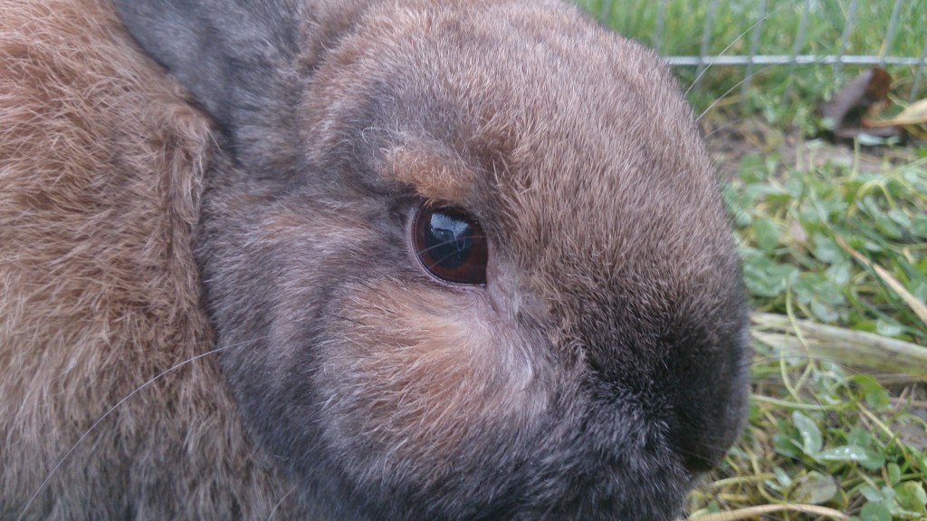
Depending on the underlying cause, treatment may be possible, for example through dental treatment. If the underlying problem is not addressed, the eye discharge is likely to return.
Antibiotic treatment is crucial. Eye drops are preferable to ointments because ointments can further block the tear duct. The tear duct can also be flushed with antibiotics. If eye drops are ineffective, a swab can be taken for an antibiogram to determine the most suitable antibiotic.
In cases of chronic widening of the tear duct, long-term intensive treatment with eye drops may be successful, often in combination with a systemic antibiotic. Mucolytics can also be used.
Good ventilation and frequent bedding changes are important to prevent ammonia build-up, and dust-free bedding should be used.
Eye Infections in Rabbits
Conjunctivitis (Inflammation of the Conjunctiva) in Rabbits
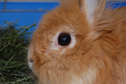
Conjunctivitis is an inflammation of the conjunctiva, the mucous membrane surrounding the eye, and is common in rabbits. It typically causes redness of the conjunctiva. Clear tear discharge is also common, while bacterial infections may cause the discharge to become pus-like.
Causes of Conjunctivitis
- Irritation
- Drafts, high levels of dust, or harmful substances in the air, such as ammonia caused by poor hygiene in the rabbit’s enclosure or smoke in smoky rooms, can irritate the eyes and lead to conjunctivitis.
- Foreign bodies, such as hay or bedding, entering the eye are another common cause.
- Allergies can also contribute to conjunctivitis.
- Injuries
- Bites or scratches can lead to conjunctivitis as a secondary condition. This form of conjunctivitis is usually unilateral, affecting only one eye, and is non-contagious.
- Infections
- Conjunctivitis can also be caused by bacterial infections, such as Pasteurella or rabbit snuffles, viruses such as Myxomatosis, or, rarely, fungi.
- When caused by an infection, conjunctivitis is usually contagious and may affect both eyes.
- Secondary Conditions
- An inflamed tear duct (dacryocystitis) often leads to conjunctivitis as a secondary condition.
Treatment
- Conjunctivitis is typically treated with eye drops or eye ointments. In cases of bacterial infection, antibiotic eye drops are often required.
- In mild cases that are not caused by injuries, Bepanthen eye ointment can be used.
- The underlying cause should be identified and addressed, and any related health conditions should also be treated.
- Corticosteroid medications should only be used if the cornea is intact, but they are generally not recommended for rabbits. Rabbits tolerate corticosteroids poorly, and they can cause liver damage, weaken the immune system, and potentially shorten lifespan.
It is important to treat both the symptoms and the underlying cause of conjunctivitis to support recovery and prevent further complications.
Meibomian Gland Disorders in Rabbits
Hypertrophy, Blockage, Meibomitis, Chalazion and Adenomas
The Meibomian glands are located on the inner side of the eyelids. Enlargement of these glands may appear as white to yellowish spots or swelling. Several different conditions can affect the Meibomian glands, most commonly inflammation of the glands (meibomitis).
When the glands become significantly thickened, a Meibomian cyst (chalazion or stye) may develop. In older rabbits, flower-like and often pigmented growths may indicate adenomas of the Meibomian glands. Pustules involving the cilial and sebaceous glands (styes) may also occur.
The exact cause is not fully understood. Possible factors include bacterial infections, genetic components and other eye diseases.
Treatment
Hypertrophy of the Meibomian glands is usually asymptomatic. However, it can occasionally contribute to inflammation around the eye or cause irritation if the enlarged glands rub against the cornea.
Regular warm compresses can help empty the glands and promote drainage. If necessary, the glands can be punctured under local anesthesia to drain the secretions. After the procedure, antibiotic eye ointments are typically applied immediately and continued for several days to prevent infection.
Eyelid Inflammation and Injury (Blepharitis) in Rabbits
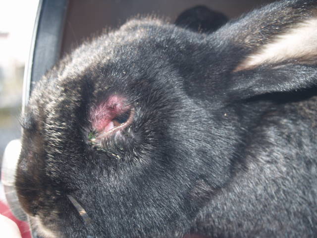
Eyelid injuries in rabbits can occur during rank fights between rabbits and often require veterinary treatment. Because the eye and surrounding tissue are very sensitive, an injury can happen quickly and without malicious intent from the other rabbit. A scratch from a claw, for example, may be enough to injure the eyelid.
A veterinarian should examine the injured rabbit eyelid and determine how severe the damage is. Depending on the extent of the injury, surgical treatment may be necessary.
It is also important to check whether the eye itself has been injured. The veterinarian can use a special dye to assess possible damage to the eye.
Antibiotic medication may be prescribed to treat the eyelid injury. Eye ointments are particularly useful because they adhere well to the eyelid.
Corticosteroid eye drops or ointments should never be used in these cases.
If an eyelid injury is left untreated, the damaged eyelid can irritate or injure the cornea.
Corneal Inflammation (Keratitis) in Rabbits
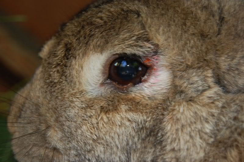
Keratitis in rabbits is an inflammation of one or more layers of the cornea, the transparent layer that covers the eye. Rabbits with corneal inflammation are often sensitive to light. Because keratitis is painful, affected rabbits may squint or keep the eye closed more often than usual.
In some cases, there may be no obvious visible changes at first. However, the cornea can develop cloudy or white spots, or the entire eye may appear to be covered by a white layer. The eye often waters, and pus may also occur. Keratitis can also be accompanied by other eye conditions, which may cause additional symptoms.
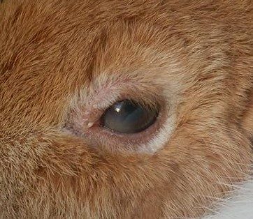
There are several possible causes of keratitis in rabbits. Viruses such as myxomatosis, fungi, and bacteria associated with rabbit snuffles can trigger corneal inflammation. Long-term untreated conjunctivitis or dacryocystitis can also lead to keratitis.
Treatment usually involves eye drops or eye ointments. Cornea-regenerating ointments or drops are also often prescribed.
Keratitis is extremely painful, even if the rabbit does not clearly show its pain. Pain relief medication must therefore be included in the treatment. If corneal inflammation is left untreated, it can lead to blindness.
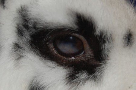
Corneal Injuries, Erosions, and Ulcerations (Ulcers) in Rabbits
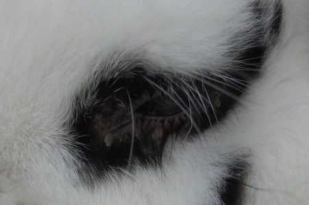
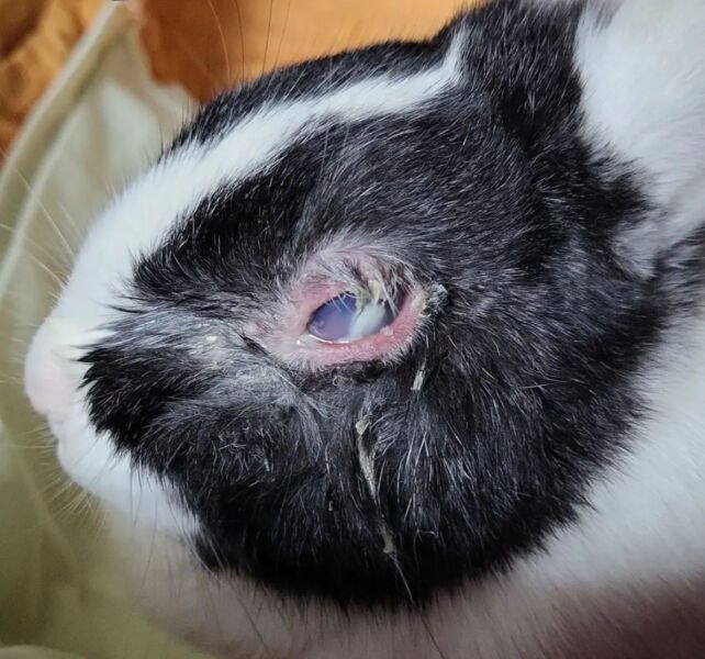
Corneal injuries and corneal ulcers in rabbits are commonly caused by eye injuries or foreign bodies. Hay stalks, dirt, thorns, twigs, or hair—especially long hair around the eyes—can injure the cornea. These injuries and foreign bodies are often not visible to the naked eye.
Another possible cause is entropion, in which the eyelid rolls inward. Dacryocystitis can also lead to corneal erosions. Drying of the eye due to protruding eyes (exophthalmos) or incomplete eyelid closure caused by Horner’s syndrome, ear infections, or other neurological causes may also result in corneal ulceration. For this reason, eyelid closure should always be tested.
To determine whether the cornea is intact or injured, a fluorescent dye is applied to the eye and the cornea is examined using a slit lamp. Corneal injuries in rabbits can be classified as superficial abrasions or erosions, perforating injuries (penetrating ulcers), or non-perforating injuries.
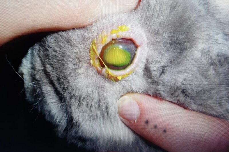
Whitish or cloudy changes in the eye should always be examined bacteriologically using a swab sample before treatment is started.
Eye injuries in rabbits heal more slowly than in dogs or cats, so patience is required. During the healing process, blood vessels grow into the injured area before complete healing occurs.
Treatment includes administering a broad-spectrum antibiotic directly into the eye together with re-epithelializing eye medications. For deeper corneal ulcers, proteinase-inhibiting eye drops such as Stromease are often used. In some cases, an artificial lens can support the healing process.
Treatment Process for Eye Injuries
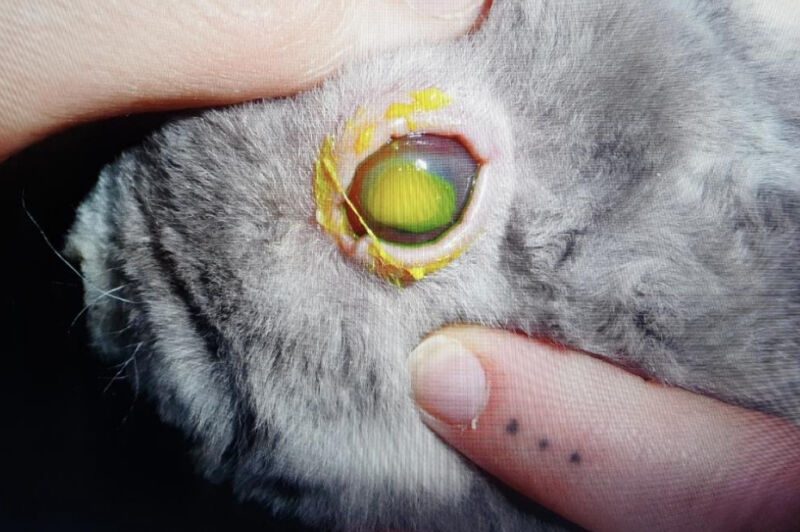
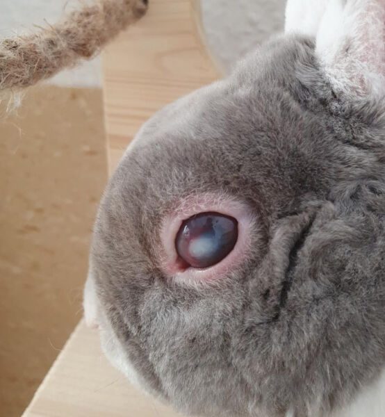
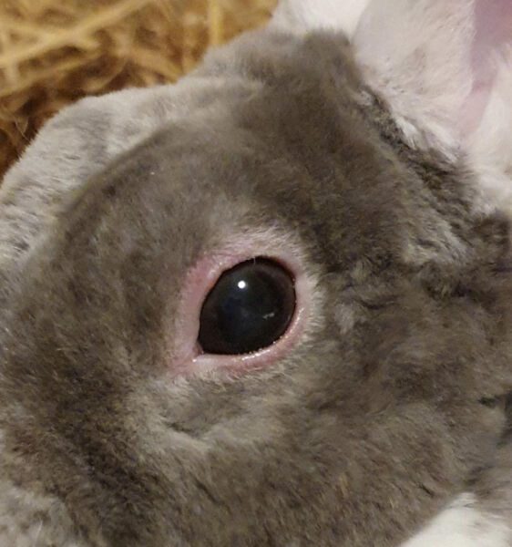
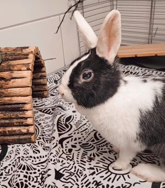
Treatment by a Veterinary Ophthalmologist with Artelac and Corneregel (2-3 Times Daily)
Entropion (Rolled Eyelid) in Rabbits
Entropion in rabbits, also known as a rolled eyelid, is an eyelid deformity in which the eyelashes rub against the surface of the eye. This causes irritation and can injure the eye.
Entropion is congenital and extremely painful because every blink causes discomfort. It is usually identified in young rabbits, and several rabbits from the same litter may be affected. Affected parent animals must be excluded from breeding programs.
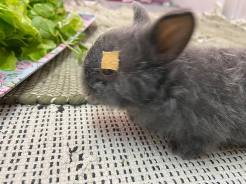
Treatment of Entropion in Rabbits
- Surgical correction: Part of the eyelid margin is removed to prevent the eyelashes from rubbing against the eye.
- Alternatively, the eyelid margin can be fixed upwards so that the rolled eyelid can heal in the correct position.
- Non-surgical correction: In some cases, taping the eyelid upwards during the growth phase can be effective, provided that it is securely fixed.
Prompt treatment is important to relieve pain and prevent further injury to the eye.
Choroiditis (Uveitis)
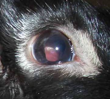
Uveitis in rabbits is a very painful inflammation of the inner eye, specifically the choroid (uvea). It is usually caused by an infection with E. cuniculi (phacoclastic uveitis), Pasteurella, or Staphylococcus. Pasteurella- or Staphylococcus-related uveitis may affect one or both eyes, while blunt trauma or a tumor usually causes unilateral uveitis.
Uveitis may appear as milky or cloudy areas in the iris or lens. Bleeding inside the eye can also cause reddish areas. One form is characterized by a yellow-white filling of the eye, while another appears as a more clearly defined, often vascularized white mass in the iris and is frequently associated with cataract formation.
Affected rabbits may blink more often than usual or squint the affected eye. The conjunctiva may be red, and the pupils may become constricted (miosis). The aqueous humor, the fluid behind the cornea, may appear cloudy. The veterinarian may also detect elevated intraocular pressure, which is painful.
Because of the pain, many affected rabbits appear unusually calm or withdrawn.
Treatment of uveitis in rabbits differs from treatment in dogs and cats because rabbits typically have a different underlying cause.
Conservative Treatment of Uveitis in Rabbits
Conservative treatment aims to prevent the progression of uveitis and address the underlying cause.
- Cortisone eye drops or eye ointments
- Liver protection: Rabbits tolerate cortisone very poorly, so liver protection is important. Milk thistle capsules or RodiCare Hepato are usually used.
- The E. cuniculi titer should be checked in the blood. In EC-positive rabbits, it is assumed that this pathogen has triggered the uveitis. In this case, the following medications are important:
- Fenbendazole (Panacur, 20 mg/kg once daily for 28 days) to help contain the pathogen
- Tetracycline-based eye ointment and a systemic antibiotic (oxytetracycline), as the pathogen is sensitive to this antibiotic
- If the intraocular pressure is too high (glaucoma), pressure-lowering eye drops are prescribed (see glaucoma).
- Atropine-containing eye drops can locally relieve pain and help prevent adhesions (synechiae). They must not be used when intraocular pressure is elevated.
- Non-steroidal anti-inflammatory eye drops may be used, such as Nepafenac (NEVANAC®), Flurbiprofen (Ocufen), Diclofenac (Voltaren), Ketorolac (Acular), or Bromfenac (Yellox).
- Anti-inflammatory pain medication, such as Meloxicam, is administered systemically. Pain relief is essential because uveitis is extremely painful.
- If these measures are not sufficient, surgical treatment must be considered because elevated intraocular pressure is very painful.
Surgical Treatment of Uveitis in Rabbits
- Phacoemulsification: This should be performed as soon as possible and can cure the uveitis.
- Eye removal (enucleation): This may be necessary if the disease is advanced and/or conservative treatment does not stop its progression.
Uveitis can lead to glaucoma, cataract, or retinal detachment.
Eye Abscess in Rabbits
Abscesses in the eye can occur in rabbits and may be mistaken for uveitis and treated incorrectly. For this reason, it is important to distinguish an eye abscess from other eye conditions.
Case Report: Corneal Abscess in a Rabbit
Conservative Treatment with Eye Preservation
“My veterinarian had also mentioned the possible removal of the eye and suspected a lens dislocation, but she referred us to Dr. Kindler in Wiesbaden, a specialist in veterinary ophthalmology.
A fluorescein test and slit lamp examination were performed there. Elvis had a pin-sized corneal injury, and the diagnosis was a corneal abscess. The affected area became increasingly white as pus formed.
We treated the eye daily with two different medications: Polyspectran and serum derived from dog blood, which was prepared directly in a centrifuge at the veterinary practice. One drop was administered in the morning, at noon, and in the evening, with a 15-minute interval between the two medications.
The veterinarian also anesthetized the eye and performed an abrasio corneae, touching the cornea to stimulate the formation of new blood vessels. Later, Elvis also received VitA POS ointment in the eye once daily.”
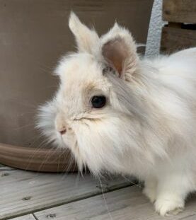
Case Study: Eye Abscess in a Rabbit
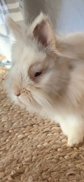
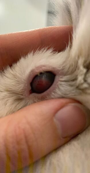
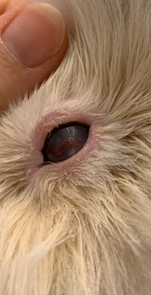
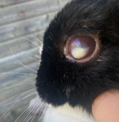
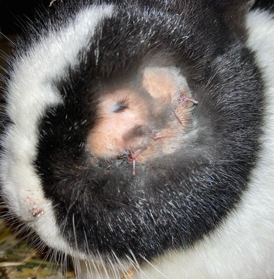
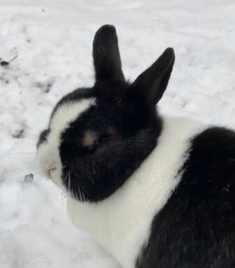
This rabbit was initially treated for uveitis. The photos show how the eye abscess gradually increased in size and significantly altered the eyeball.
By the time the abscess was correctly diagnosed, the condition had progressed so far that the eye had to be removed. After the eye removal, the rabbit recovered very well and was finally pain-free.
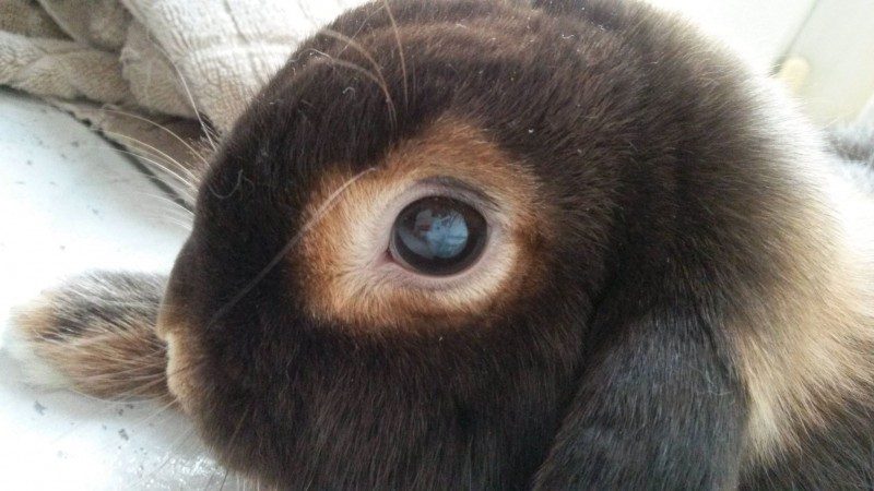
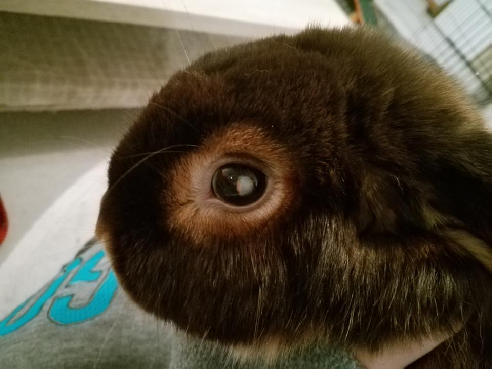
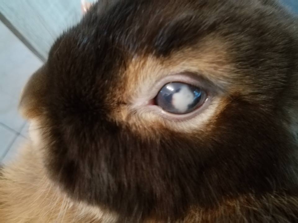
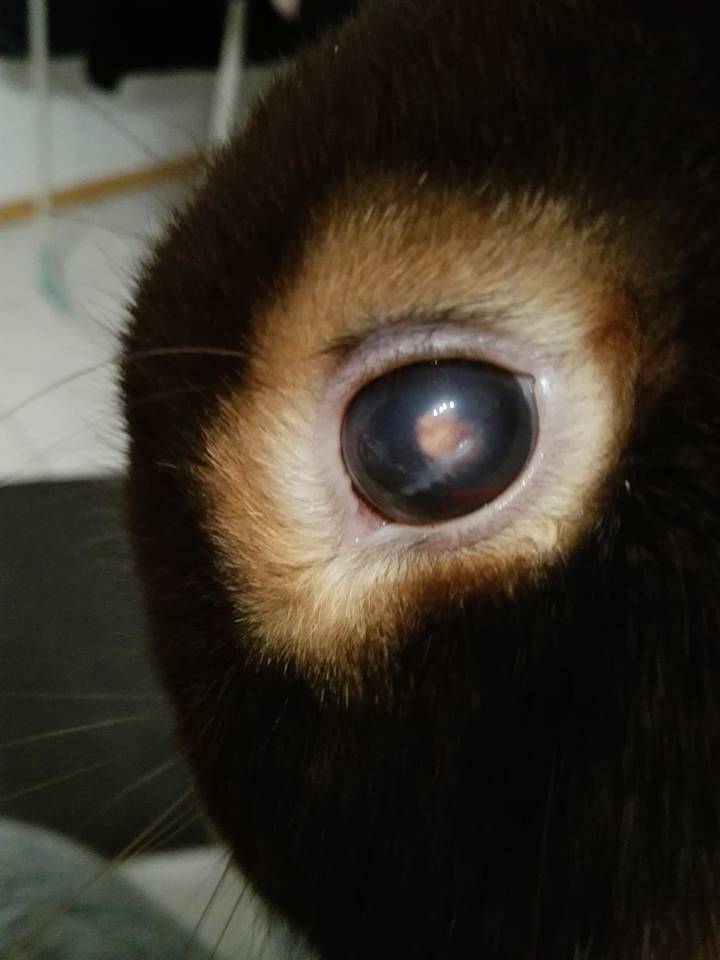
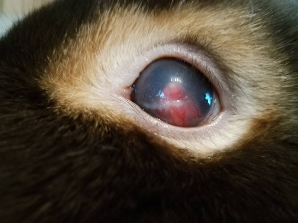
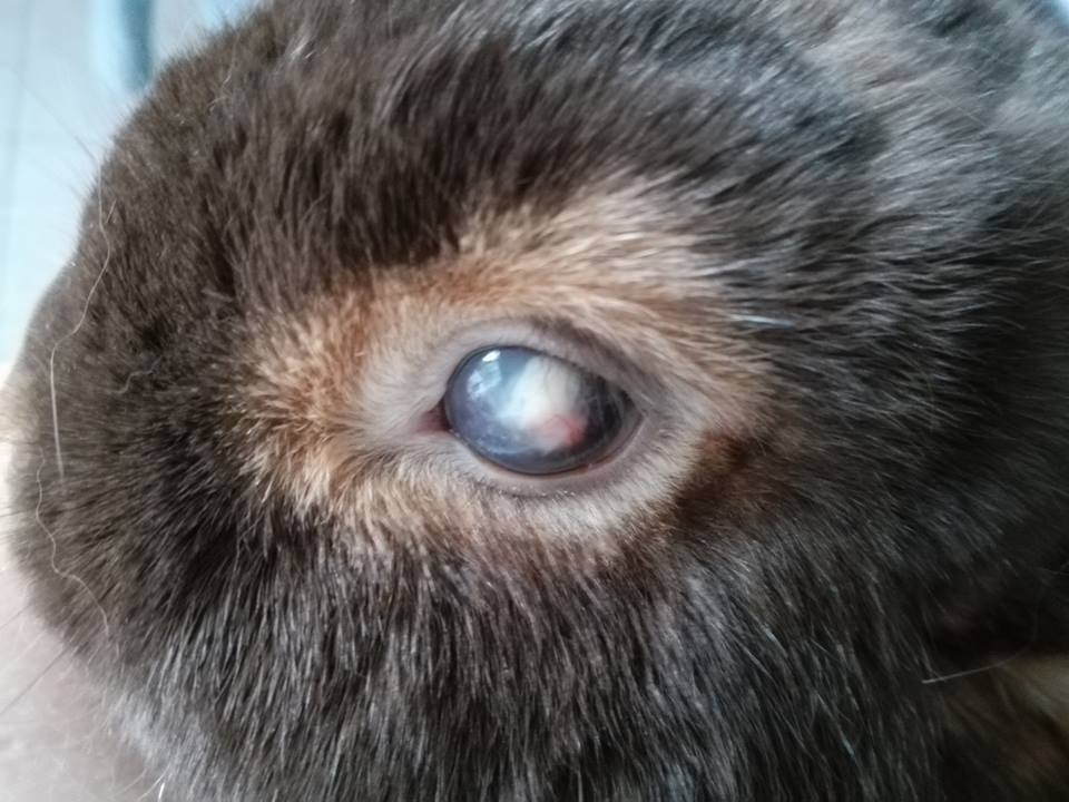
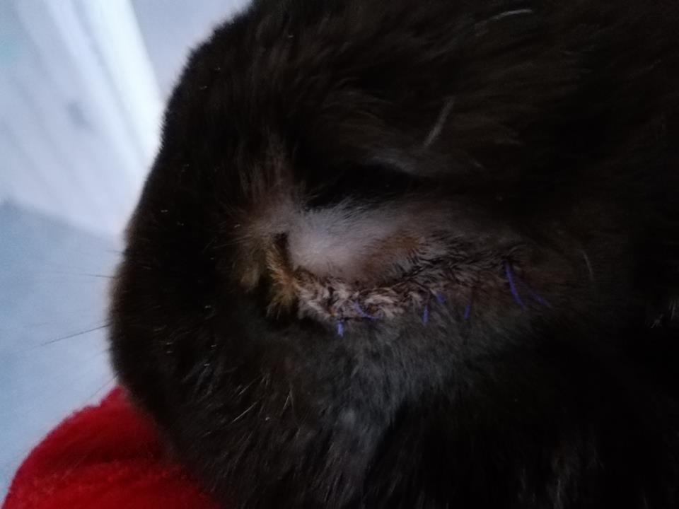
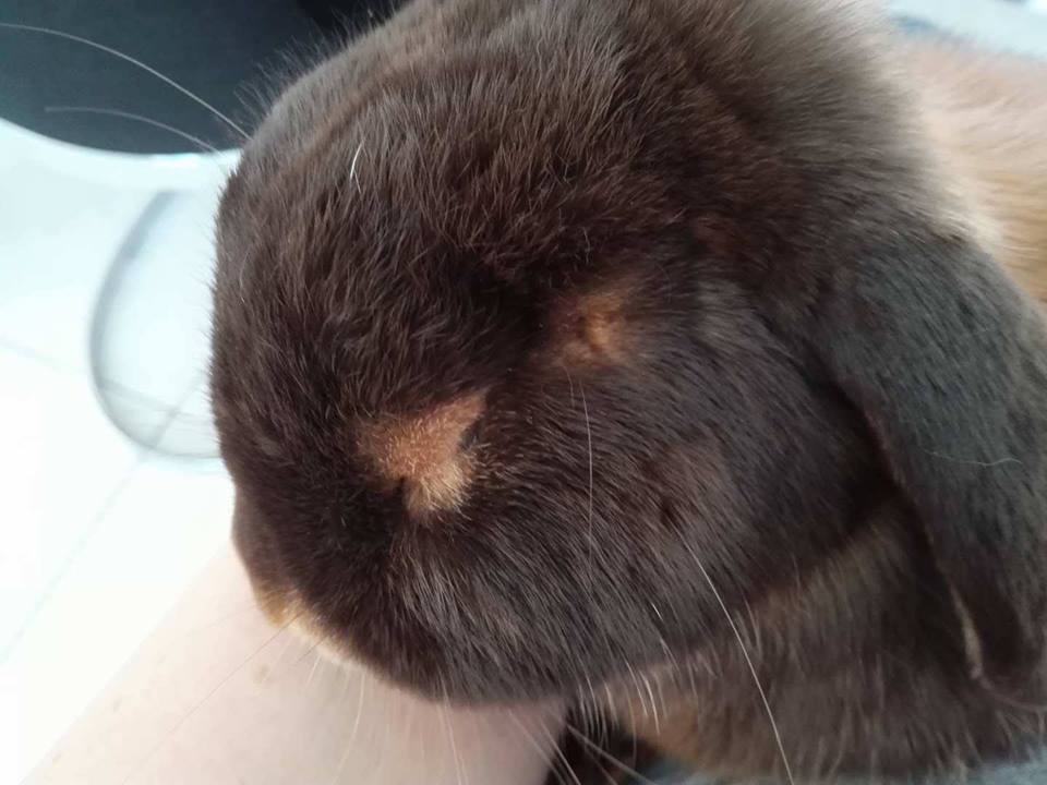
Maja, 8 years old, developed a corneal abscess following an eye injury, which later progressed to an intraocular abscess. Her eye had to be removed, but she recovered very well and is doing great.
Maja’s case shows that rabbits can still have a good outcome even at an advanced age.
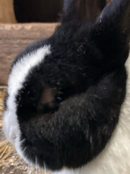
Pseudopterygium and Pterygium in Rabbits
Pseudopterygium in rabbits is a growth of the conjunctiva that is believed to be inherited. As long as the conjunctiva is not fused with the eye and does not cause problems, treatment is not necessary.
If the conjunctiva becomes fused with the cornea (pterygium), surgical treatment is recommended.
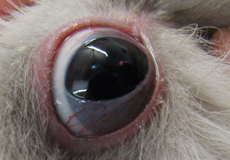
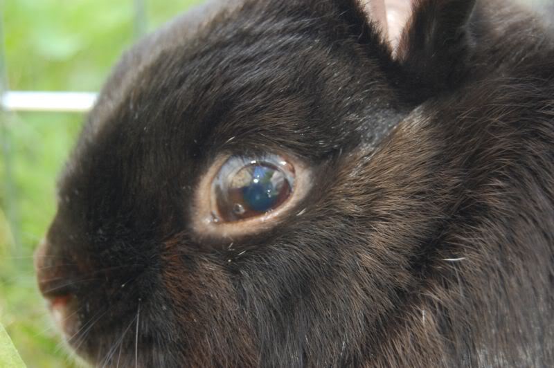
Cataracts (Lens Opacity) in Rabbits
Cataracts in rabbits are caused by an opacity of the lens. They may be hereditary or develop slowly as an age-related condition, but they are most commonly caused by E. cuniculi.
Diabetes can also cause cataracts, although this is extremely rare in rabbits.
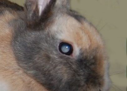
Case Report: Cataracts and Glaucoma in a Rabbit
Maple developed cataracts due to E. cuniculi. Both eyes later developed glaucoma and had to be removed in separate surgeries, about one year apart.
Despite being blind, Maple manages wonderfully and is completely happy and pain-free.
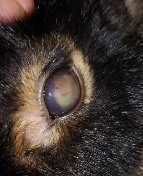
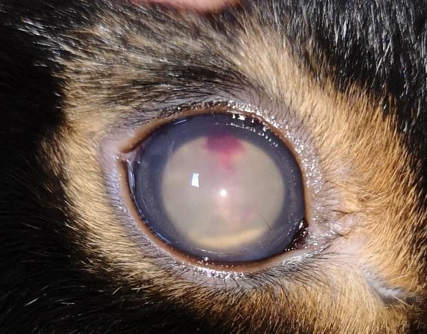
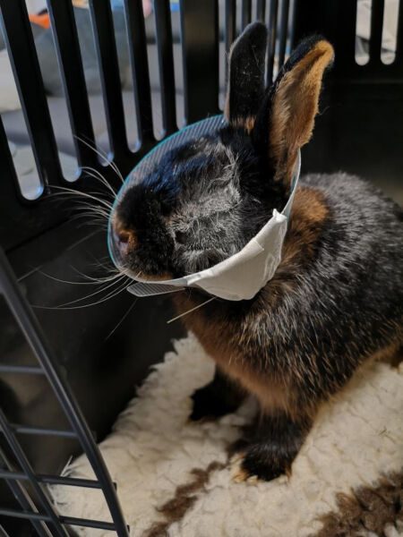
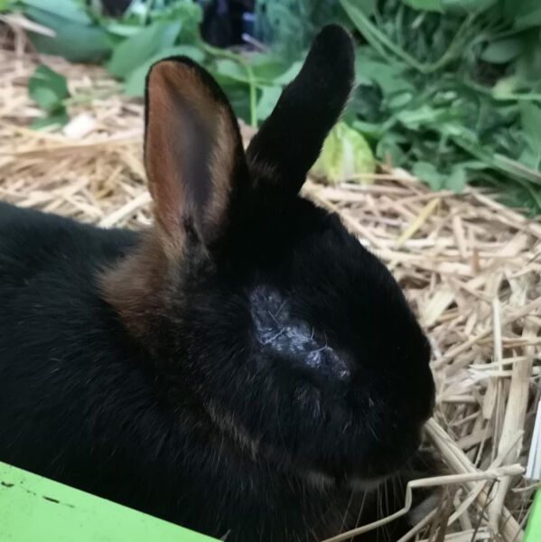
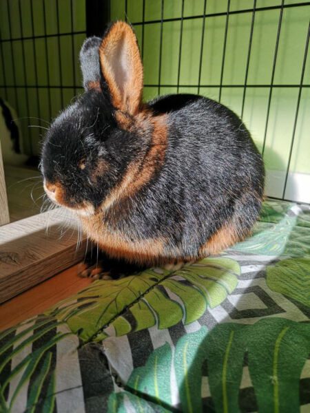
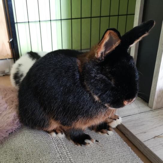
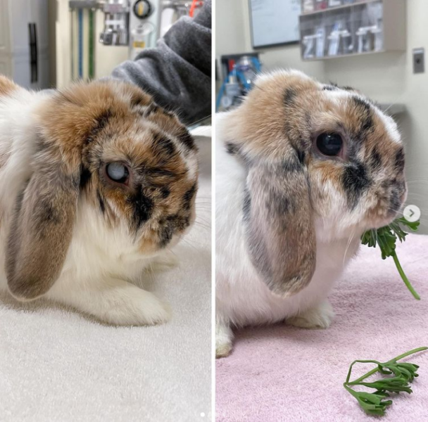
Photo: Nicola Di Girolamo, Instagram: @nic_the.animal.doctor
Glaucoma in Rabbits (Increased Intraocular Pressure)
Glaucoma in rabbits is associated with increased intraocular pressure and can cause the eyeball to become enlarged or more prominent. The condition can appear in different ways.
Affected rabbits may squint, blink more frequently, or become sensitive to light. The eye may appear reddish due to increased blood circulation, and the pupil is often enlarged. If the eyeball becomes enlarged, the eyelids may no longer close completely. Areas of the eye that are no longer fully covered by the eyelids can then develop a gray-white layer.
Glaucoma is very painful, although rabbits rarely show obvious signs of pain. Instead, they may become quieter, withdrawn, or apathetic.
In rabbits, glaucoma is usually caused by a pre-existing condition such as uveitis. Hereditary glaucoma has been described in White New Zealand rabbits, but it can also occur in other white rabbits due to autosomal recessive inherited goniodysplasia.
The intraocular pressure rises significantly because of an imbalance in the circulation of the aqueous humor. Normally, the aqueous humor circulates through the angle of the anterior chamber between the front and back parts of the eye. If the canal in the chamber angle is blocked, the aqueous humor cannot drain properly, resulting in increased intraocular pressure. Elevated intraocular pressure can also occur if too much aqueous humor is produced.
Treatment of Glaucoma in Rabbits
In the early stages, medications are used to reduce the intraocular pressure. In later stages, lowering the pressure may no longer be easily possible, and the eye may need to be removed to relieve the pain.
Untreated glaucoma can lead to blindness. However, treatment is still necessary after blindness has occurred because the affected rabbit continues to experience severe pain.
Pain is rarely indicated by squinting alone and is more often reflected in behavioral changes such as withdrawal or apathetic behavior.
EC-positive rabbits should receive Panacur concurrently to reduce the pathogen load.
A veterinary ophthalmologist will carefully examine the glaucoma and prescribe the appropriate medications.
Pharmacological Treatment
- Treating the underlying cause: The E. cuniculi titer should be determined because E. cuniculi may be the cause. In EC-positive rabbits, Panacur should also be administered.
- Inhibition of aqueous humor production:
- Dorzolamide (Trusopt)
- Brinzolamide (Azopt)
- Dorzolamide/Timolol (Cosopt)
- Brinzolamide/Timolol (Azarga)
- Beta-blockers are generally not used in rabbits:
- Timolol maleate
- Levobunolol
- Betaxolol
- Improvement of aqueous humor drainage:
- Miotics cause pupil constriction to improve drainage. They cause contraction of the ciliary muscle and increase vascular permeability. Depending on the type of glaucoma, they may not always be suitable.
- Direct-acting miotics:
- Pilocarpine
- Indirect-acting miotics:
- Demecarium bromide
- Echothiophate iodide
- Improvement of uveoscleral aqueous humor drainage:
- Latanoprost (Xalatan)
- Travoprost (Travatan)
- Tafluprost (Taflotan)
- Unoprostone (Rescula)
- Amlodipine: Calcium channel blockers increase blood flow and reduce vascular resistance.
- Reduction of intraocular volume:
The use of mannitol is now considered outdated. - Anti-inflammatory pain medication:
- Meloxicam
Surgical Treatment of Glaucoma in Rabbits
Depending on how advanced the glaucoma is, different surgical treatment options are available. A veterinary ophthalmologist can advise on the most appropriate approach.
Exophthalmos (Protruding Eye) in Rabbits
Exophthalmos in rabbits means that the eye protrudes from the eye socket. It can affect one eye (unilateral exophthalmos) or both eyes (bilateral exophthalmos).
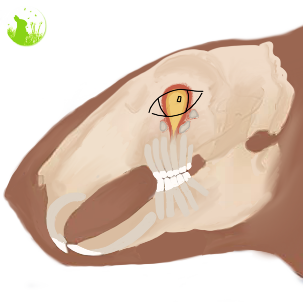
A protruding eye in rabbits is most commonly caused by dental disease that leads to a granuloma or abscess at the tooth roots behind the eye. In extremely rare cases, a tumor behind the eye, such as lymphoma of the Harderian glands, may be responsible.
Other possible causes include injuries such as blunt trauma, hematomas, tear duct diseases, fat prolapse, foreign bodies, or a salivary gland cyst. In one reported case, the eye protruded because of a cyst containing a tapeworm larva (Taenia serialis).
Bilateral Exophthalmos in Rabbits
When both eyes protrude, the cause is usually increased blood pressure, which causes the blood vessels behind the eyes to swell and push the eyes forward.
This can occur with heart disease or masses in front of the heart, such as thymoma, lymphoma, or abscesses. Thymoma is particularly notable in connection with bilateral exophthalmos. Thrombosis of both jugular veins can also cause the eyes to protrude.
Excessive fat deposits in the eye sockets of overweight rabbits can also contribute to bilateral exophthalmos.
Rabbits with protruding eyes often show reduced food intake and behavioral changes, including withdrawal. The affected eye may also partially dry out if the eyelids can no longer close completely.
Treatment of Exophthalmos in Rabbits
The treatment of exophthalmos in rabbits depends on the underlying cause.
Unilateral Protruding Eye
If only one eye is protruding, a computed tomography (CT) scan should be performed if possible to identify the underlying cause and allow targeted treatment.
The most common cause is dental disease. This can be diagnosed using a CT scan or X-rays in four planes, together with a thorough examination of the oral cavity. The upper molars, usually P3–M3, are typically affected.
If a jaw abscess is located behind the eye, the affected tooth must be removed by a veterinary dentist and the abscess surgically drained. In most cases, the eye can be preserved. The rabbit will also receive an effective antibiotic and pain relief.
Some less specialized veterinarians may remove the entire eye, but this approach is now considered outdated.
Tumors are extremely rare. If a tumor is present, it must be surgically removed. In many cases, the eye also needs to be removed during surgery. However, rabbits can generally continue to live comfortably in their group, even in enclosures with multiple levels.
Bilateral Protruding Eyes
In rabbits with bilateral exophthalmos, heart disease should be ruled out. An echocardiogram, ECG, and blood pressure measurement are recommended to detect heart disease as well as abscesses, lymphomas, or thymomas around the heart.
In some cases, these conditions can also be detected by X-ray. Treatment can then be started according to the underlying condition.
A protruding eye is extremely painful and can lead to drying of the eye due to incomplete eyelid closure, as well as circulatory failure. The condition should therefore be treated as soon as possible. Strong pain relief is necessary until surgery.
Eyeball Removals and Eye-Saving Surgeries
Case Reports
Schnee (4 years old)
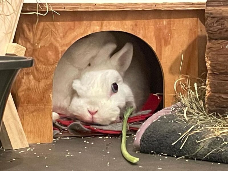
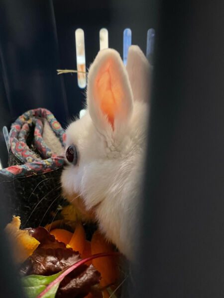
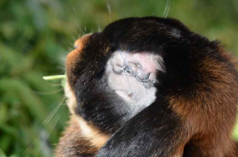
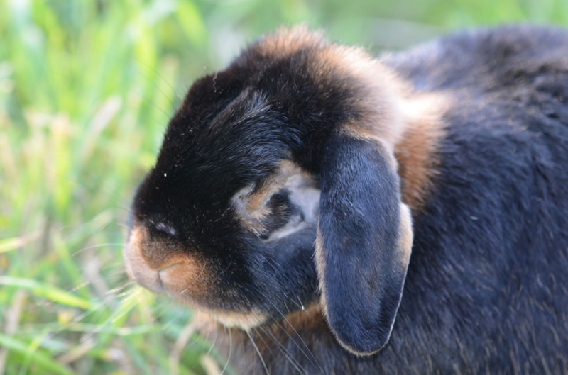
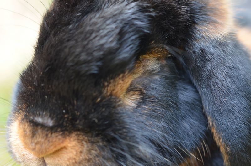
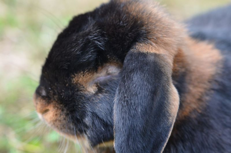
The eye was protruding, and the rabbit could no longer close it. The triggering tooth was removed, and the eye was preserved. In the second-to-last photo, you can see him the day after surgery; he can now close his eye again.
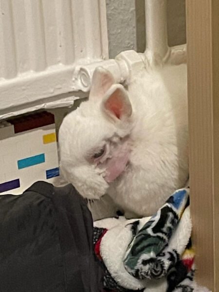
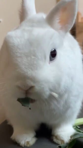
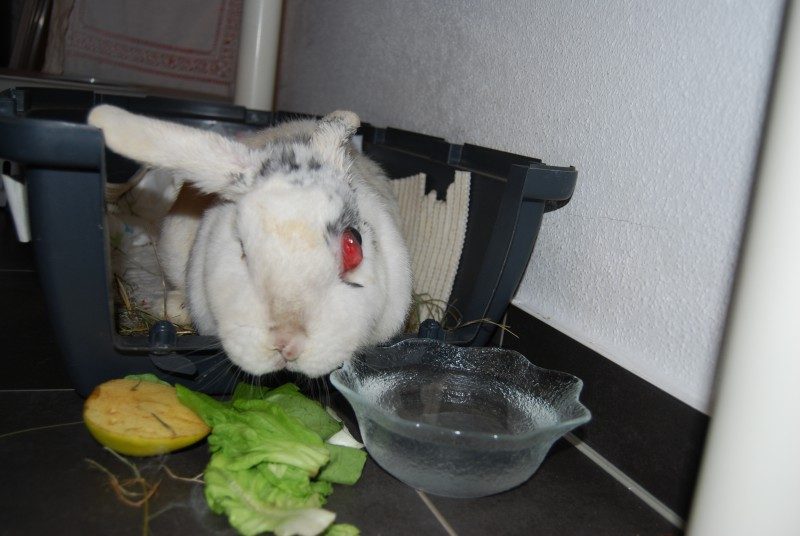
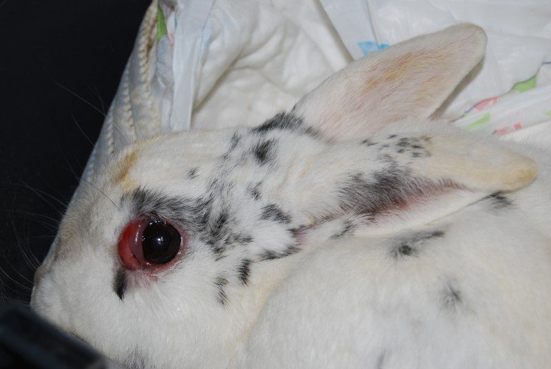
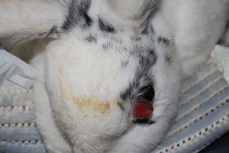
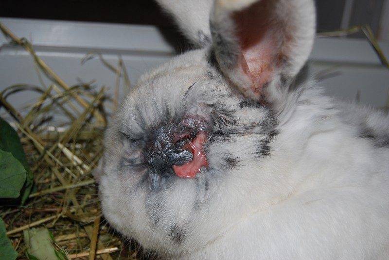
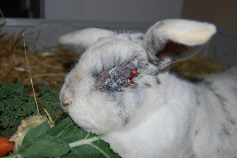
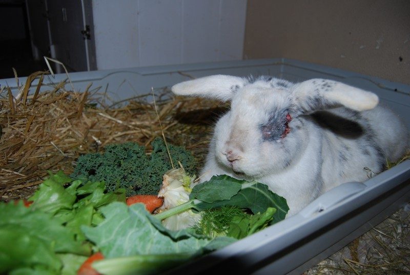
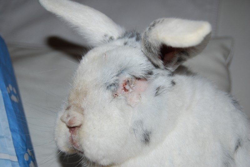
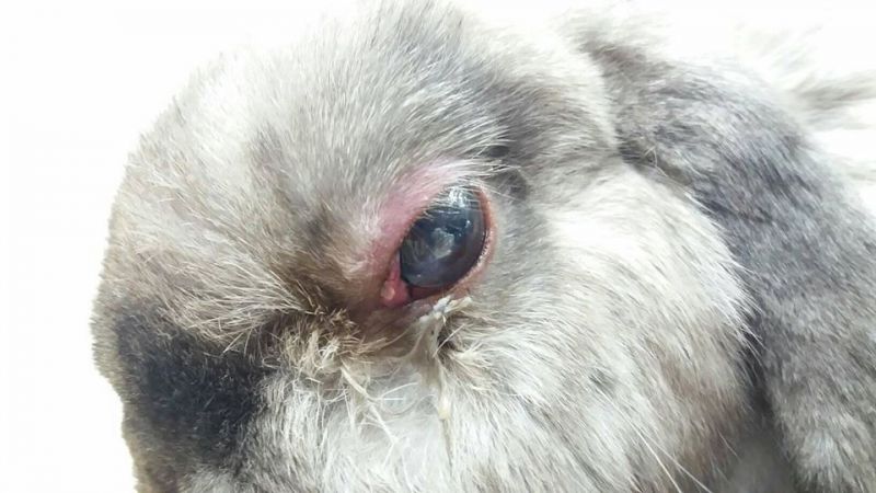
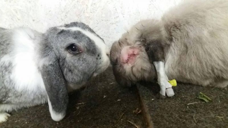
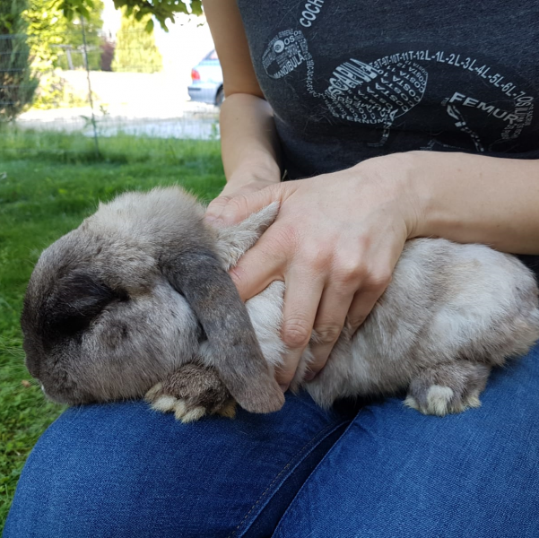
Ernie was operated on at over 8 years old. After the surgery, he immediately felt much better because the severe pain was gone.
Third Eyelid Prolapse (Haw’s Gland Hyperplasia) in Rabbits
The third eyelid in rabbits, also known as the nictitating membrane or nictitating gland, is normally not visible. An exception can occur in some large meat rabbit breeds, where it may be visible more often.
If the third eyelid becomes visible temporarily or permanently, it can indicate a health problem. It may also become more noticeable during stress, heat, or when the rabbit lowers its head.
Possible causes of third eyelid prolapse in rabbits include injury or trauma to the third eyelid, enlargement near the heart such as thymoma or lymphoma, associated high blood pressure, heart disease, infections such as rabbit snuffles, eye diseases, and abscesses or tumors behind the eye.
Because there are many possible causes, identifying the underlying problem can be difficult. A thorough examination should include an echocardiogram and a detailed eye examination. In some cases, a dental examination may also be recommended.
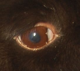
Fat Eye (Fatty Eye) in Rabbits
Fat eye in rabbits is an eye condition characterized by a sagging eyelid and a protruding conjunctival sac beneath the eye. The condition is hereditary and is particularly pronounced in certain rabbit breeds, including German Giants and German Giant Spotted rabbits.
Unfortunately, fat eye is often unintentionally bred into these rabbits because the condition usually does not develop until several months of age, around puberty. Rabbits with fat eyes should therefore be excluded from breeding programs.
In most cases, fat eye does not require treatment.
Commonly Used Eye Medications in Rabbits
Antibiotic Eye Ointments and Drops
Examples include Cephenicol CA, Floxal, Chibroxin, Refobacin, Posifenicol, Gent Ophtal, and Oxytetracycline.
These are used for bacterial infections of the eye or surrounding area. Chloramphenicol, such as Cephenicol CA, is considered the most effective antibiotic for eye infections. Infections of the nasolacrimal duct are treated with antibiotic eye drops rather than ointments.
Eye drops and ointments can be swallowed through the nasolacrimal duct and must therefore be safe for ingestion (follow the PLACE rule).
Eye ointments remain in the eye longer and usually need to be applied less frequently. Eye drops are generally easier to administer and do not clog the nasolacrimal duct. Some rabbits are sensitive to eye ointments; in these cases, drops are preferred.
Corticosteroid Eye Ointments and Drops
Examples include Cephenidex CA/DEX, Ultracortenol, and Isopto-Max.
These are generally unsuitable for most diseases in rabbits, with the exception of uveitis. They can shorten the rabbit’s lifespan, affect the immune system, and negatively impact the liver. Some rabbits react more strongly than others, while others tolerate them well.
Corticosteroid eye medications are used for non-infectious inflammation of the eye or surrounding area. They should never be used in cases of corneal injury because they can interfere with healing.
Pain-Relieving Eye Drops
Non-steroidal anti-inflammatory eye drops, such as Nepafenac (NEVANAC®), are used to relieve pain and inflammation in the eye.
Regenerative Eye Ointments and Drops
Products such as Corneregel and Regepithel are typically used for corneal defects.
Moisturizing Eye Drops
These are used when the eye does not produce sufficient tears or when the eyelids cannot close completely.
Vitamin A-Containing Eye Ointments and Drops
Products such as Vitagel are used to promote healing and regeneration of the eye’s surface.
Atropine-Containing Eye Drops
These are used for several purposes, including pupil dilation, pain relief, and the prevention of adhesions.
Eye Drops to Lower Intraocular Pressure
Examples include Azopt, Travatan, Xalatan, and Azarga. These medications are used to lower intraocular pressure, for example in rabbits with glaucoma.
Bepanthen Eye and Nose Ointment
Used for disorders of the skin and mucous membranes.
Homeopathic Eye Drops
Common options include Echinacea and Euphrasia eye drops. Euphrasia is used for inflammation, while Echinacea is used to regulate fluid balance in the eye.
About This Article
Original content by Viola Schillinger, veterinarian
Veterinarian specializing in rabbit health, nutrition and species-appropriate husbandry, and founder of Kaninchenwiese.de, a long-established German rabbit care and health resource reaching around 500,000 visitors per month.English translation and adaptation by Erika Kühne Escobar
Medically reviewed by Viola Schillinger, veterinarian
Last medically reviewed: August 2026
Sources include:
Böhmer, E. (2011): Zahnheilkunde bei Kaninchen und Nagern: Lehrbuch und Atlas; mit 27 Tabellen. Schattauer.
Butler, K., DeGeorge, B., Dunn, G., VanGyzen, J., Gruaz, M., van Praag, A., & van Praag, E. (2016): Heterochromia of the iris in rabbits belonging to the Dutch breed.
Eckert, Y., Ferkau, A., & Thöle, M. (2022): Augenerkrankungen beim Kaninchen. Der praktische Tierarzt, 103(4), 352-372.
Ewringmann, A. (2016): Leitsymptome beim Kaninchen: diagnostischer Leitfaden und Therapie; Georg Thieme Verlag.
Khelik, I., Johnson III, J. G., McCool, E., Fentiman, K., Soler, E., Tully, T., & Brandão, J. (2023): Aberrant conjunctival overgrowth with corneal adhesions in two pet rabbits (Oryctolagus cuniculus). Journal of Exotic Pet Medicine, 46, 32-37.
Gabriel S. (2016): Praxisbuch Zahnmedizin beim Heimtier. Stuttgart: Enke
Glöckner, B. (2016): Dacryocystitis beim Kaninchen. team. konkret, 12(04), 8-13.
Künzel, Frank (2013): Thymom – ein unterdiagnostiziertes klinisches Problem beim Kaninchen? Veterinär-Spiegel 23.01: 22-25.
Miyake H, Oda T, Katsuta O, Seno M, Nakamura M. A (2017): Novel Model of Meibomian Gland Dysfunction Induced with Complete Freund’s Adjuvant in Rabbits. Vision. 2017; 1(1):10
Millar TJ, Pearson ML. (2002): The effects of dietary and pharmacological manipulation on lipid production in the meibomian and harderian glands of the rabbit. Adv Exp Med Biol. 2002:431-40.
Morrisey, J. K., & McEntee, M. (2005): [Englisch] Therapeutic Options for Thymoma in the Rabbits
O’Reilly, A., McCowan, C., Hardman, C., & Stanley, R. (2002): Taenia serialis causing exophthalmos in a pet rabbit. Veterinary ophthalmology, 5(3), 227-230.
van Praag, E.: Meibomian cyst or the growth of a white nodule on the rabbit eyelid. [http://www.medirabbit.com/EN/Eye_diseases/Disorder/Meib/Meib_en.htm; Stand: 22.03.2023]
Rother, N.; Lazarz, B. (2018): Hyperplasie der Meibom´schen Drüsen beim Kleinsäuger. Kleintiermedizin 18(06); 254-257
Spiess, A (2023): Untersuchungsgang und häufige Erkrankungen am Kaninchenauge. Kleine Heimtiere
Walde, I. (Ed.). (2008): Augenheilkunde: Lehrbuch und Atlas; Hund, Katze, Kaninchen und Meerschweinchen. Schattauer Verlag.
Williams, D. (2012): Rabbit and rodent ophthalmology – Medirabbit [http://www.medirabbit.com/EN/Eye_diseases/Eyes_diseases_rabbit.pdf; abgerufen am 21.03.2023]
Zinke, J. (2004): Ganzheitliche Behandlung von Kaninchen und Meerschweinchen: Anatomie, Pathologie, Praxiserfahrungen; Georg Thieme Verlag.