Rabbit dental disease is one of the most common health problems in rabbits. It is often detected too late and can severely affect a rabbit’s overall health, leading to serious secondary conditions.
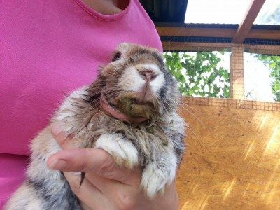
Warning: Rabbits with dental problems can slowly starve — even when food is always available.
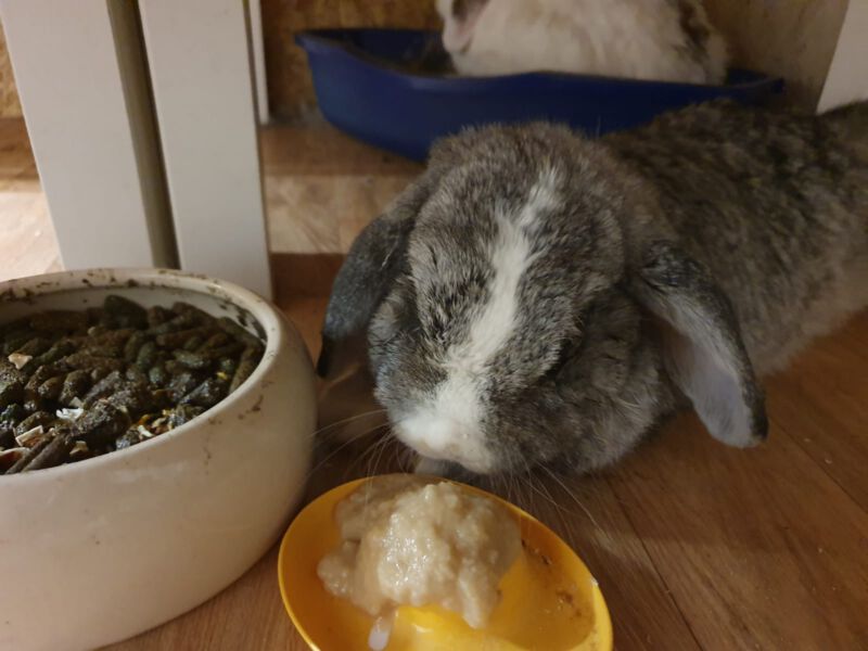
Dental misalignment or tooth pain makes it difficult or impossible for them to eat properly. Many rabbits will still approach their food, pick at it, and appear to be eating — but in reality, they are not consuming enough to survive
Contents
- Symptoms of Rabbit Dental Disease: How to Recognize the Signs
- Common Symptoms of Rabbit Dental Disease
- Advanced Signs of Rabbit Dental Disease
- Causes of Rabbit Dental Disease: Why Do Dental Problems Develop?
- Insufficient Tooth Wear Due to an Improper Diet
- Diet as the main cause
- What rabbits actually need
- Why chewing matters
- Food That Is Too Hard Can Damage Teeth
- Why this is harmful
- Other Causes of Rabbit Dental Disease
- Genetic and Congenital Causes
- Improper Diet During Growth
- Trauma and Tooth Injuries
- Age-Related Dental Changes
- Mineral Deficiencies and Metabolic Disorders
- Reduced Food Intake
- Ear Infections and Changed Chewing Behavior
- Hand-Raising of Rabbits
- Expert Insight: Why Diet Is the Main Cause of Rabbit Dental Disease
- Why modern feeding causes dental problems
- The myth of “hard food”
- Rabbits are built for soft, fibrous foods
- Important conclusion
- Why Domestic Rabbits Develop Dental Disease More Often Than Wild Rabbits
- Differences between wild and domestic rabbits
- How diet changes jaw development
- Why hay-heavy diets can increase risk
- Why dried food is not a solution
- What rabbits actually need
- Scientific conclusion
- Examination and Diagnosis of Rabbit Dental Disease
- Finding a Rabbit-Savvy Veterinarian
- Examining the Molars (Back Teeth)
- ⚠️ Important: Avoid Mouth Spreaders
- Diagnostic Tools for Rabbit Dental Disease
- Oral Examination
- ⚠️ Mouth Spreaders – Use With Caution
- X-Rays: Essential for Diagnosis
- Sedation and Positioning
- Tear Duct Diagnostics
- Advanced Imaging
- Treatment of Rabbit Dental Disease
- Trimming Overgrown Incisors
- Removal of Severely Misaligned Incisors
- Broken Incisors
- Incisor Misalignment in Young Rabbits
- Surgical Removal of Infected Teeth
- Treatment of Molar (Back Teeth) Diseases in Rabbits
- Grinding of Overgrown Molars
- Follow-Up and X-Rays
- Surgical Removal of Diseased Molars
- Do Opposing Teeth Need to Be Removed?
- Tooth Growth After Removal
- Retrograde Tooth Growth
- Treatment of Oral Injuries
- Dental Treatment With or Without Anesthesia
- Why anesthesia is important
- ⚠️ Risks of treatment without anesthesia
- ⚠️ Mouth spreaders must not be used on conscious rabbits
- Why proper dental tools matter
- Benefits of treatment under anesthesia
- When treatment without anesthesia may be considered
- Important conclusion
- Common Mistake: Dental Treatment Without Anesthesia
- Why “tooth clipping” is a problem
- Why early proper treatment matters
- Real-life outcomes
- Safety of sedation
- ⚠️ Why clipping is dangerous
- Important conclusion
- Case ReportsMoritz, a Satin Rabbit with Numerous Jaw Abscesses and Dental Problems
- Nutrition for Dental Diseases
- Feeding After Dental Surgeries and for General Dental Diseases
Symptoms of Rabbit Dental Disease: How to Recognize the Signs
The symptoms of rabbit dental disease can vary widely. In many cases, rabbits show only subtle signs, and often just one or two symptoms appear at the same time — even when a serious dental problem is already present.
Because of this, dental issues in rabbits are frequently overlooked until they become severe.
Common Symptoms of Rabbit Dental Disease
Rabbits with dental disease often show subtle but serious signs. Even one of the following symptoms can indicate a dental problem:
- Weight loss or emaciation due to reduced food intake
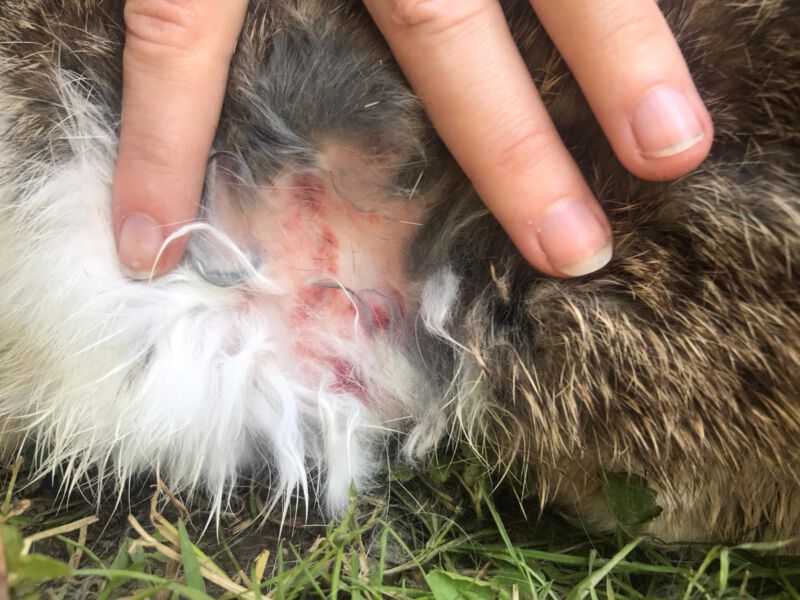
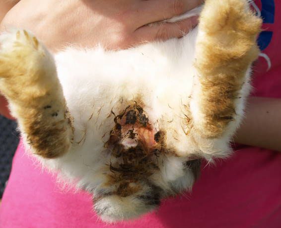
- Wet chin or drooling (saliva on the mouth, chin, neck, or front paws)
- Digestive problems, such as diarrhea, bloating, or irregular/mushy poops
- Teeth grinding (often a sign of pain)
- Overgrown, broken, or misaligned front teeth
- Reduced fecal output or “stringy” poops connected by fibers
- Difficulty closing the mouth or holding it slightly open
- Selective eating (avoiding hard foods, preferring soft or shredded food)
- Difficulty eating (food falls out of the mouth or cannot be chewed properly)
- Slower eating combined with constant hunger or food searching
- Chewing only on one side of the mouth
- Increased water intake or excessive drinking
Even mild or occasional symptoms should be taken seriously, as rabbit dental disease often progresses unnoticed.
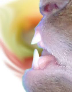
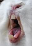
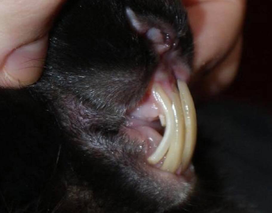

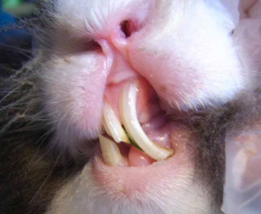

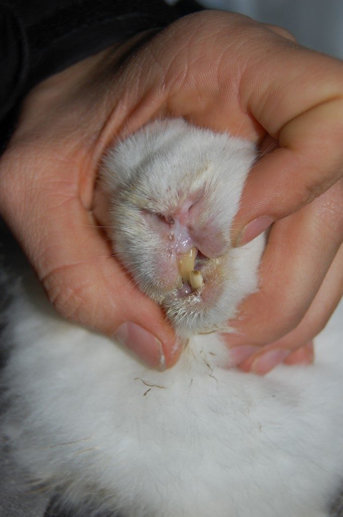
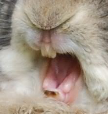

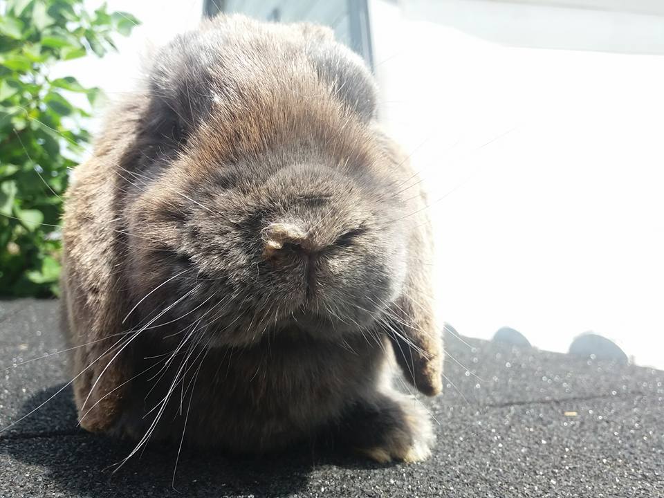
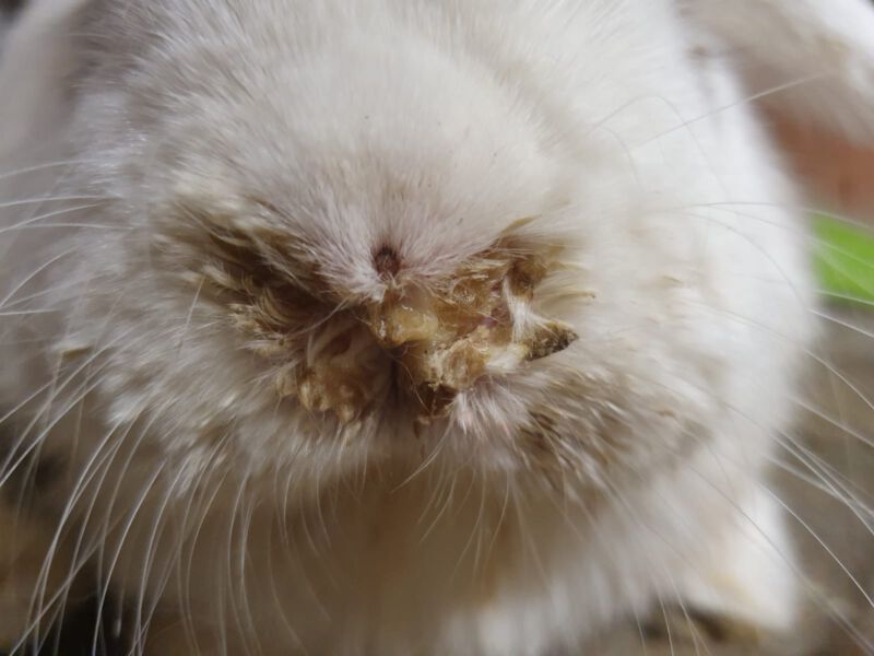
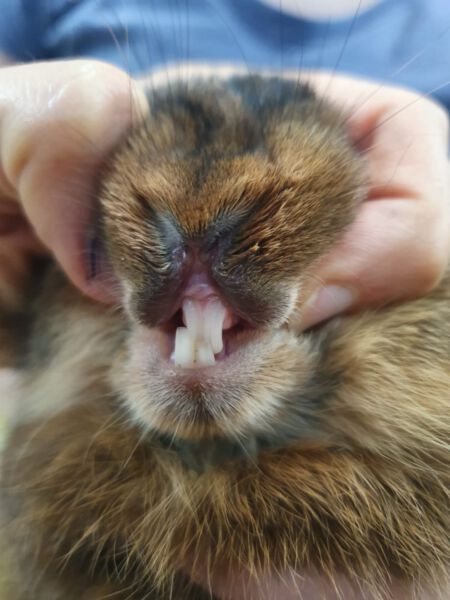

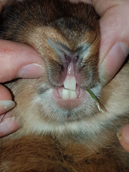

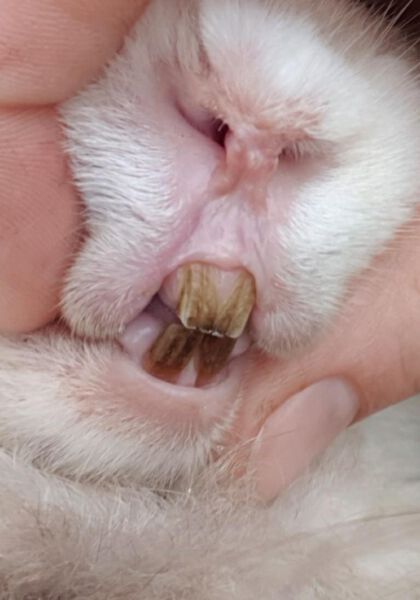
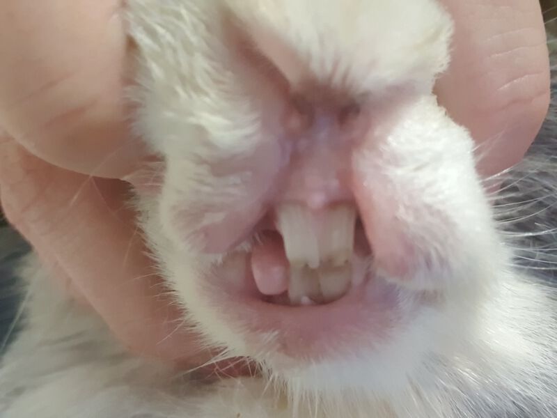
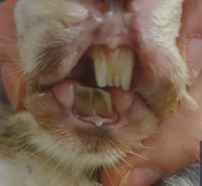
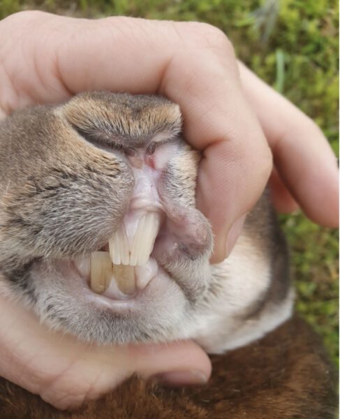
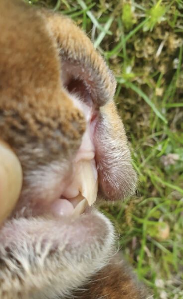
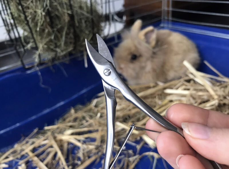
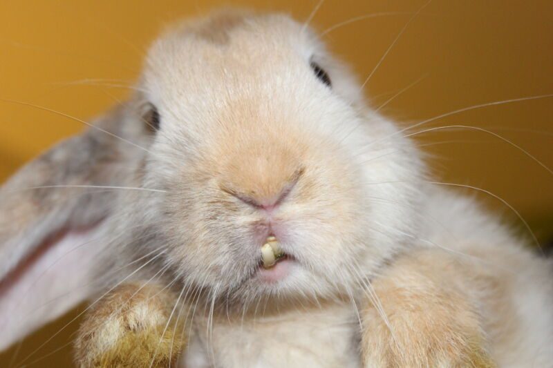
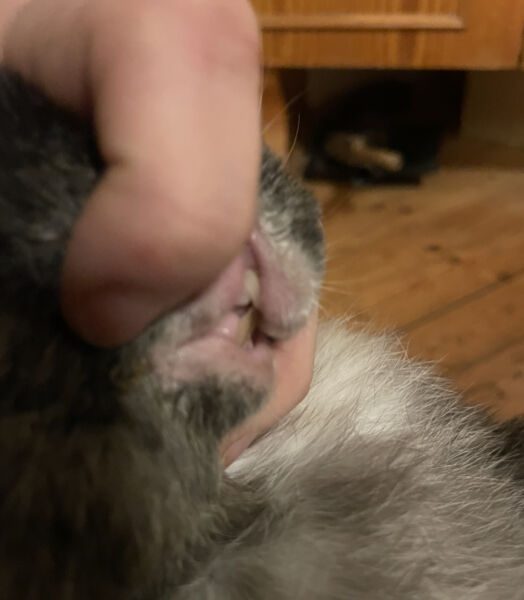
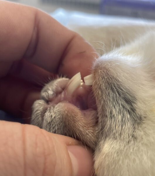
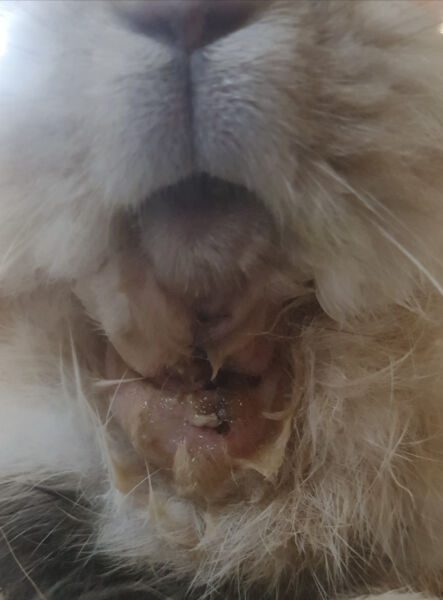
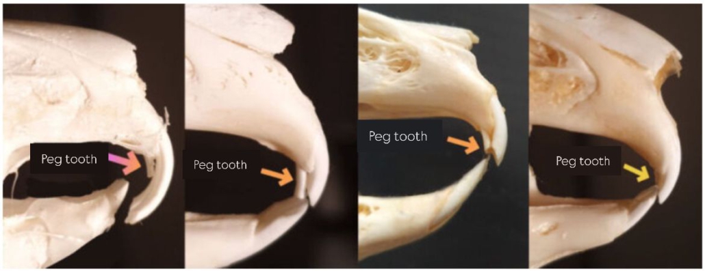
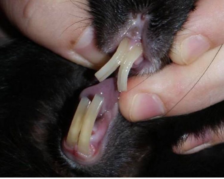
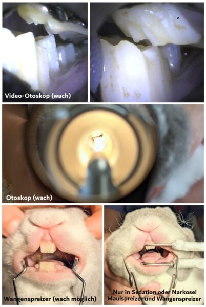
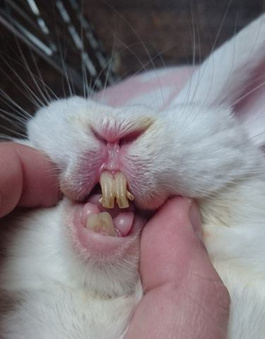
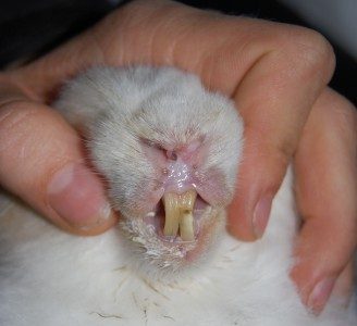
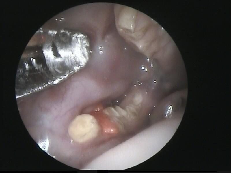
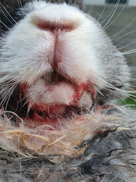
The peg teeth can be examined from the side during a health check. The three images on the left show an unhealthy set of teeth with overgrown peg teeth. It’s important to check not only the length but also the shape of the surface. This surface should extend the cutting edge of the incisors, running horizontally or slightly sloping toward the molars, but it should not form a triangular shape (as seen in image 3). The image on the right shows a healthy set of teeth, where the peg teeth fit snugly against the cutting surface of the incisors. Additionally, the length of the incisors should be assessed, ensuring the upper and lower incisors are approximately the same length.
There are also indirect symptoms that are often not immediately linked to dental diseases and, as a result, frequently go unnoticed or are only recognized after multiple veterinary visits and proper diagnostics:
Advanced Signs of Rabbit Dental Disease
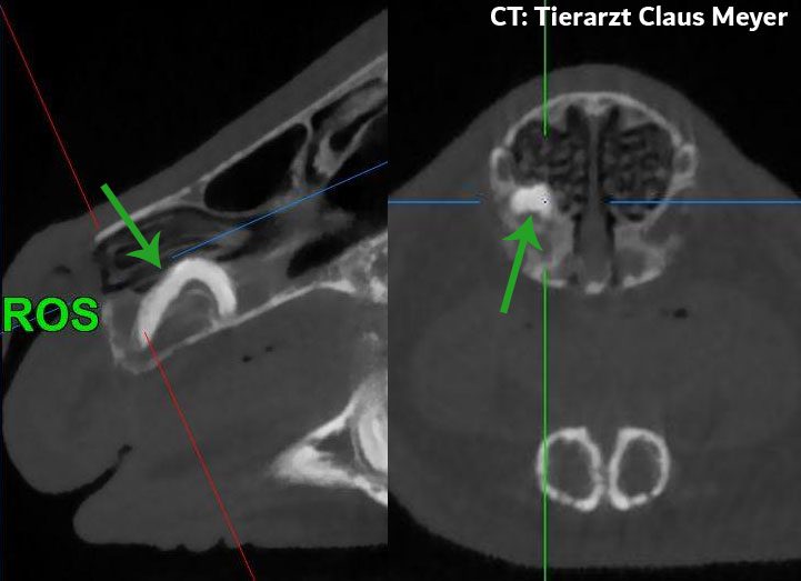
In more advanced stages of rabbit dental disease, problems often develop deep in the jaw and may affect surrounding structures.
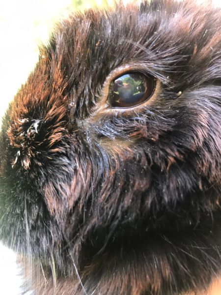
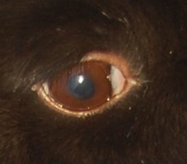
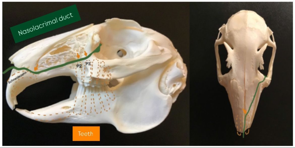
One important area is the nasolacrimal duct, which runs very close to the tooth roots. When teeth grow abnormally or become overgrown, they can press on this duct and cause visible symptoms.
Common advanced symptoms include:
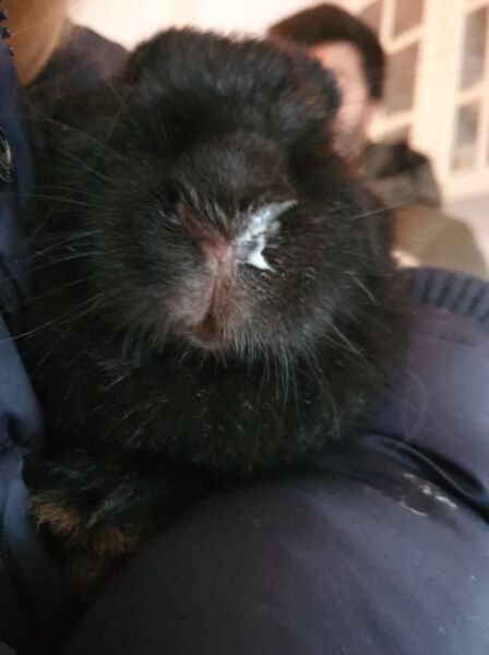
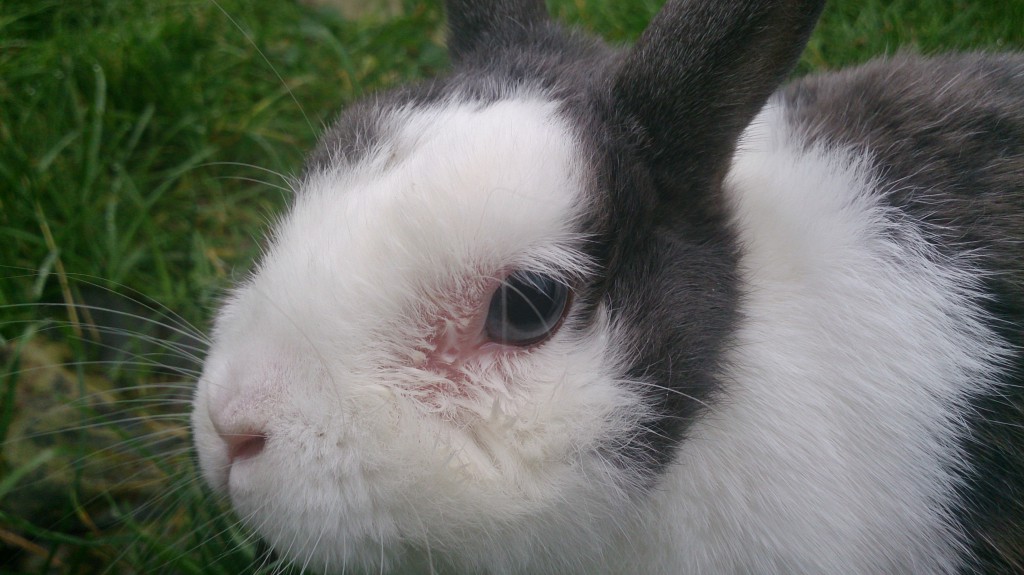
- Eye discharge (one-sided or both eyes)
→ caused by pressure on the nasolacrimal duct - Protruding eye (bulging eye)
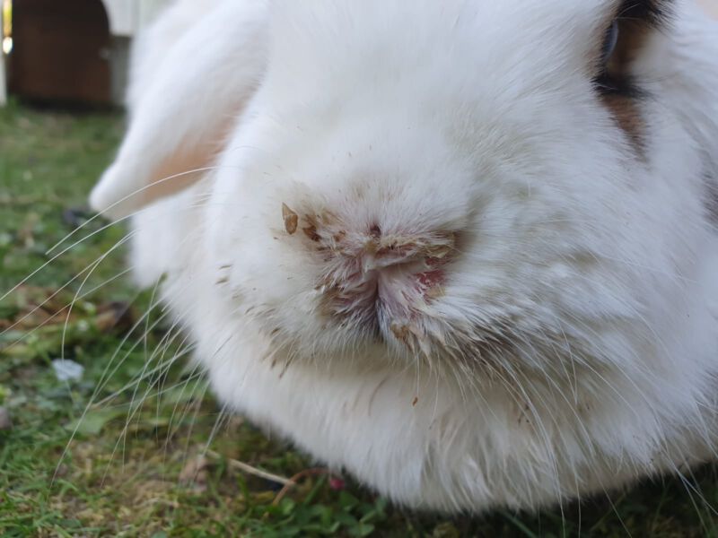
→ often linked to severe dental root problems - Nasal discharge (“snuffles”)
→ due to inflammation or blocked tear ducts - Lumps along the jaw
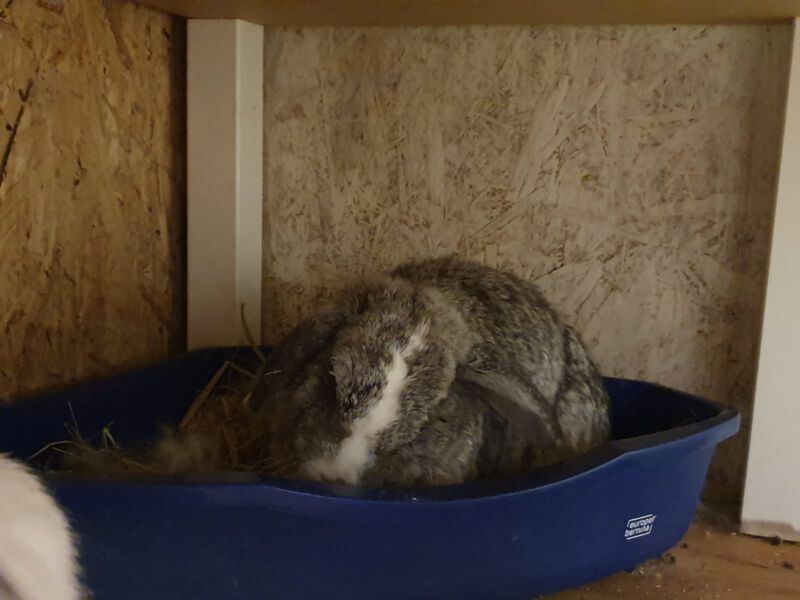
→ can indicate abscesses or infections in the jawbone - Reduced activity and withdrawal
→ rabbits become quieter and less active due to pain
Causes of Rabbit Dental Disease: Why Do Dental Problems Develop?
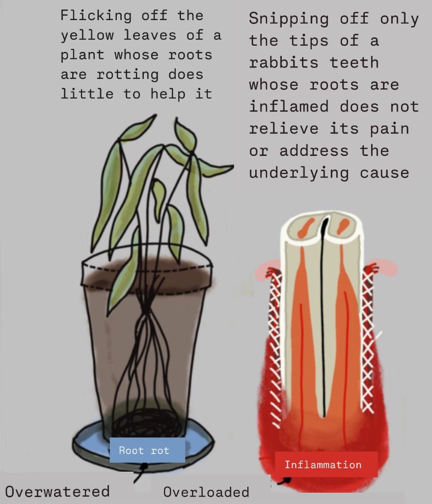
Rabbit dental disease is most
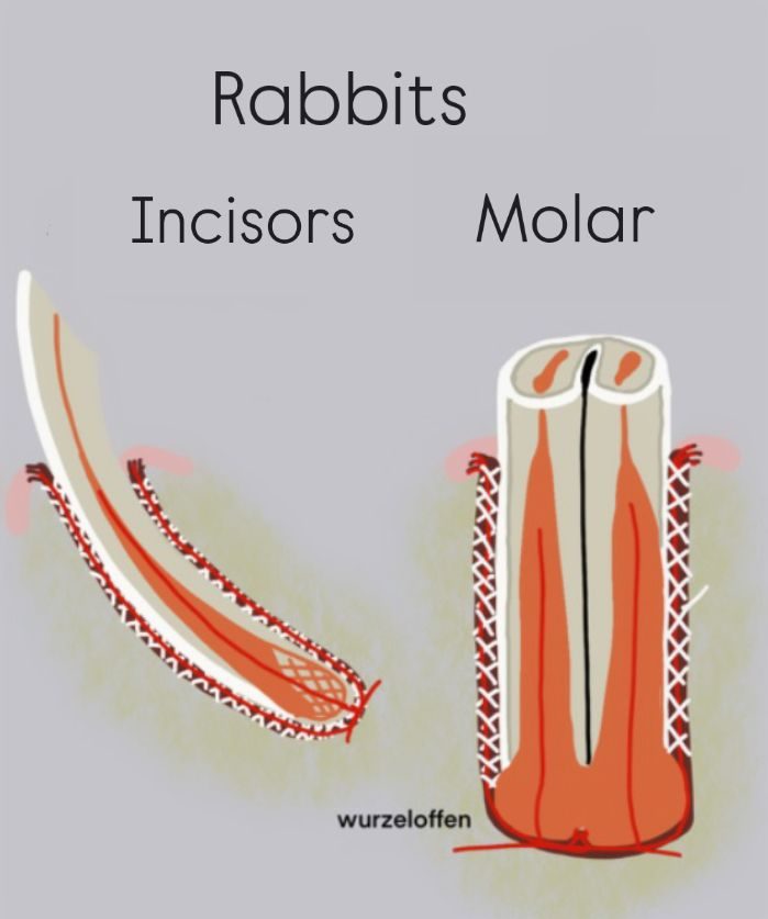
Rabbits do not have tooth roots because new material is constantly formed at the bottom of their ever-growing teeth, and the specialized tooth-supporting apparatus pushes them upwards, causing them to grow. This delicate tooth-supporting system, with its periodontal ligaments, can be damaged by improper food, preventing the teeth from being pushed upwards. As a result, the tooth „grows“ into the bone and often into the eye socket, the tear duct, or it may break through the lower jaw outward.
Insufficient Tooth Wear Due to an Improper Diet
The most common cause of rabbit dental disease is insufficient tooth wear, which is directly linked to an improper diet.
Rabbit teeth grow continuously throughout their lives. This means they must be worn down naturally through constant chewing.
Diet as the main cause
Feeding the wrong types of food is one of the leading causes of dental problems in rabbits.
❌ Problematic foods include:
- dry food (pellets)
- bread
- grains (corn, seeds, etc.)
- energy-rich foods
These foods do not provide proper tooth wear and can quickly lead to dental issues.
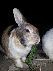
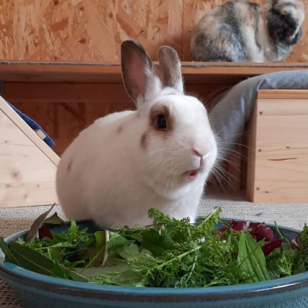
What rabbits actually need
For healthy teeth, rabbits require a diet based on:
- fibrous, structured foods (not ground or processed)
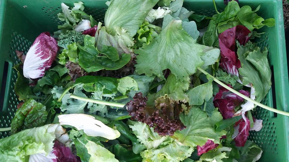
- fresh, leafy greens
- herbs and wild plants
- variety and constant availability
Examples include:
- grass
- dandelion
- clover
- leafy branches
- vegetable greens
- leafy vegetables
The more a rabbit chews, the better the natural tooth wear.
Why chewing matters
Rabbits achieve proper tooth wear when they:
- chew frequently
- chew fibrous food
- eat foods that require grinding (not biting)
Constant chewing is essential for preventing dental disease.
Food That Is Too Hard Can Damage Teeth
Feeding foods that are too hard can also cause serious dental problems.
❌ Examples:
- dry food
- hard bread
- mainly solid vegetables or fruits
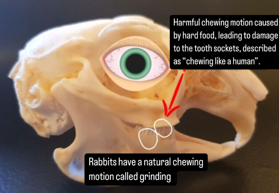
Why this is harmful
Rabbits process these foods differently:
- they break them apart (like humans)
- instead of grinding them (natural chewing motion)
This leads to:
- excessive pressure on tooth roots
- strain on the jaw
- long-term dental damage
Over time, this can result in:
- retrograde tooth growth
- inflammation
- serious dental disease
Other Causes of Rabbit Dental Disease
In addition to diet-related issues, there are several other causes of rabbit dental disease that should not be overlooked.
Genetic and Congenital Causes
Some rabbits are born with dental problems due to genetics.
- Jaw misalignments (e.g. in dwarf rabbits with short skulls)
- Genetic tooth malformations (e.g. Satin rabbits)
Poor breeding practices significantly increase the risk.
Improper Diet During Growth
Rabbits raised on an unnatural diet may develop long-term dental issues.
- feeding mainly pellets, dry food, and hay
- lack of natural chewing during development
This can affect jaw shape and muscle development, leading to dental disease later in life.
Trauma and Tooth Injuries
Physical damage to the teeth can cause serious dental problems.
Common causes include:
- biting on cage bars
- falls or collisions (e.g. running into walls)
- improper handling (e.g. mouth spreaders)
- clipping teeth with pliers (“nipping”)
These injuries can lead to misalignment, fractures, or root damage.
Age-Related Dental Changes
As rabbits age, their teeth can shift naturally.
This can lead to:
- uneven wear
- misalignment
- increased risk of dental disease
Mineral Deficiencies and Metabolic Disorders
Dental problems can also result from nutritional imbalances.
Possible causes:
- calcium deficiency
- vitamin D deficiency (especially in indoor rabbits)
- disrupted calcium-phosphorus balance
- medications or other illnesses
Vitamin D (“sun vitamin”) is especially important for indoor rabbits.
Reduced Food Intake
If a rabbit eats less due to illness:
- chewing decreases
- tooth wear is reduced
This quickly leads to dental problems.
Ear Infections and Changed Chewing Behavior
Undiagnosed ear infections can affect chewing.
- rabbits may chew differently or less
- often seen in certain breeds (e.g. dwarf rabbits)
Hand-Raising of Rabbits
- mineral deficiencies during growth
- improper tooth development
Hand-raised rabbits may develop:
This increases the risk of dental disease later in life.
Expert Insight: Why Diet Is the Main Cause of Rabbit Dental Disease
According to veterinarian Dr. med. vet. Diana Ruf, improper feeding is the main cause of dental problems in rabbits.
Rabbits have continuously growing teeth. Even if they eat less or chew less, their teeth will continue to grow.
Why modern feeding causes dental problems
When rabbits are fed energy-rich foods (such as pellets or dry food), they feel full faster and chew less.
Less chewing = insufficient tooth wear
As a result, the teeth become too long and dental disease develops.
The myth of “hard food”
A common misconception is that hard foods like dry bread help wear down teeth.
This is incorrect.
Rabbit teeth are not worn down by hard food —
they are worn down by tooth-to-tooth contact during natural chewing (grinding).
Rabbits are built for soft, fibrous foods
Rabbit teeth are highly specialized for:
- grinding fresh greens
- chewing fibrous, leafy plants
If they are fed inappropriate food that cannot be properly ground:
- the chewing motion changes
- the tooth roots are stressed incorrectly
- serious dental problems can develop
Important conclusion
- Most dental problems in rabbits are caused by improper diet.
- “Misalignments” are rarely congenital —
they are usually the result of insufficient tooth wear and incorrect chewing stress.
Source:
Dr. med. vet. Diana Ruf http://tieraerztin-ruf.de/2017/07/24/gesunde-ernaehrung-von-meerschweinchen-und-kaninchen/
Why Domestic Rabbits Develop Dental Disease More Often Than Wild Rabbits
Domestic rabbits are far more likely to develop rabbit dental disease than their wild counterparts — and diet plays a major role in this difference.
Differences between wild and domestic rabbits
Studies show that the teeth themselves are almost identical in wild and domestic rabbits.
The key difference lies in the skull shape and chewing mechanics:
- Domestic rabbits
- shorter, higher skull
- steeper chewing muscles
- higher pressure on the back molars
- Wild rabbits
- longer, flatter skull
- more even distribution of chewing force
This means domestic rabbits experience more pressure on specific teeth, increasing the risk of dental problems.
How diet changes jaw development
The type of food rabbits eat — especially at a young age — directly affects how their jaw develops.
This is known as phenotypic plasticity.
- Rabbits fed coarse, unnatural food (hay-heavy, pellets)
→ develop stronger jaw muscles
→ create higher chewing pressureCombined with unnatural chewing motion, this can lead to:
- retrograde tooth root growth
- tooth root damage
- chronic dental disease
Why hay-heavy diets can increase risk
Although hay is important, a hay-heavy diet can create problems:
- prolonged chewing → increased pressure on teeth
- risk of gum irritation from coarse plant material
Why dried food is not a solution
Replacing fresh food with dried herbs or “crumbly” food is not effective.
These foods:
- break apart too easily
- do not create proper tooth wear
What rabbits actually need
Rabbits require a diet that supports their natural chewing motion:
- fresh, soft, leafy foods
- food that can be cut and ground sideways (natural grinding motion)
Examples:
- fresh meadow plants (summer)
- leafy vegetables (winter)
Hay should always be available —
but it should not be the main part of the diet.
Scientific conclusion
- Diet directly affects jaw development, chewing mechanics, and long-term dental health.
- Most dental problems in domestic rabbits are linked to unnatural feeding and chewing patterns.
Source:
Fr. Dr. Böhmer: „Unique Specialization of the Rabbit’s Dentition: Are There Differences Between Wild and Domestic Rabbits?“ Rodentia Nager & Co.
Examination and Diagnosis of Rabbit Dental Disease
Rabbit dental disease can be difficult to detect and requires a careful and experienced examination.
While many veterinarians can identify basic dental issues, proper diagnosis and treatment often require a rabbit-savvy veterinarian or a specialist in rabbit dentistry.

Finding a Rabbit-Savvy Veterinarian
An experienced veterinarian can usually examine the incisors (front teeth) quite easily.
They will check:
- color
- shape
- length
- alignment
If the incisors are overgrown or misaligned, the molars are almost always affected as well.
Examining the Molars (Back Teeth)
The molars are much more difficult to assess, as they are located deep inside the mouth.
For an initial examination, veterinarians may use:
- an otoscope
- or a cheek retractor
These tools allow a basic evaluation without causing unnecessary stress.
⚠️ Important: Avoid Mouth Spreaders
A mouth spreader should not be used in rabbits.
It can cause serious injuries, such as:
- jaw fractures
- loose or damaged teeth
- dental misalignment
- tooth root infections and abscesses
In addition, the stress and pain can cause rabbits to panic, which may lead to:
- injuries to the spine
- injuries to the hind legs
Safe handling and proper tools are essential when diagnosing rabbit dental disease.
Caution: Please do not give pea flakes, nuts, parsnips, or any other white food before the appointment (e.g., to lure the rabbit into the box), as it could be mistaken for pus during the oral examination.
Diagnostic Tools for Rabbit Dental Disease
Accurate diagnosis of rabbit dental disease requires more than a simple visual check, as most dental problems are hidden deep in the tooth roots.
Oral Examination
The best way to examine the oral cavity of an awake rabbit is by using a video otoscope, which provides a detailed view of the teeth and mouth.
Basic examinations can also be performed using:
- an otoscope
- a cheek retractor
- a headlamp for better visibility
These tools allow an initial assessment without causing unnecessary stress.
⚠️ Mouth Spreaders – Use With Caution
Mouth spreaders are attached to the front teeth and should only be used under sedation or anesthesia.
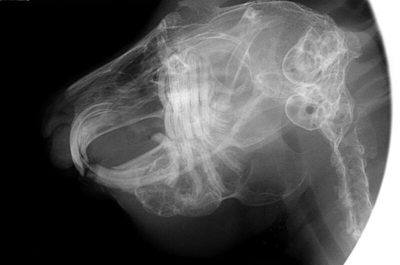
X-Rays: Essential for Diagnosis
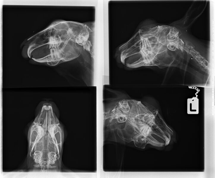
If rabbit dental disease is suspected, X-rays are essential.
- Around 80% of dental problems are located in the tooth roots or jaw, making them invisible from the outside.
For an accurate diagnosis, X-rays are taken from multiple angles:
- Lateral view → evaluates tooth shape and root length
- 45-degree angled view → allows detailed assessment of individual tooth roots
These views help detect:
- root elongation
- abscesses
- misalignment
Evaluation is often based on established reference lines (e.g. Böhmer & Crossley).
Sedation and Positioning
X-rays can often be taken without sedation.
However:
Sedation or anesthesia may be used to:
- improve positioning
- obtain clearer images
- allow additional imaging angles
Tear Duct Diagnostics
If the tear duct is affected, further diagnostics may include:
- flushing the tear duct (to check if it is blocked)
- staining the eye (to detect corneal damage or inflammation)
- contrast X-rays (to identify narrowing or obstruction)
Advanced Imaging
In some cases, more advanced imaging is required:
- intraoral X-rays
- CT scans
- MRI scans
These methods provide more detailed information, especially for:
- tooth roots
- abscesses
- tear duct involvement
Treatment of Rabbit Dental Disease
The treatment of rabbit dental disease depends on the severity and type of the dental problem. Early diagnosis is essential to prevent serious complications.
Incisor Tooth Diseases
Changes or misalignments of the incisor teeth are almost always caused by molar diseases! The teeth must be X-rayed in multiple planes by a dental specialist for rabbits and examined under anesthesia in the mouth, with appropriate treatment if necessary. Simply shortening the incisors without thoroughly examining/treating the molars does not solve the problem!
Trimming Overgrown Incisors
Overgrown incisors must be carefully trimmed according to proper dental reference lines.
Important:
- Teeth should never be clipped with pliers
- Only rotating dental tools should be used
Clipping teeth can cause:
- microfractures
- loose or unstable teeth
- abnormal tooth growth
- increased risk of abscesses and infections
In some cases, trimming can be performed without anesthesia.
However, trimming often needs to be repeated every 3–6 weeks, which is why permanent solutions may be considered.
Removal of Severely Misaligned Incisors
If the incisors are severely misaligned and cannot function properly:
👉 surgical removal may be the best option
After removal:
- rabbits cannot bite food
- food must be chopped or shredded
This can significantly improve quality of life and reduce repeated veterinary visits.
Broken Incisors
Broken incisors usually regrow naturally.
However:
- the opposing tooth may overgrow and must be monitored or trimmed
- the underlying cause must be identified
Possible causes include:
- root infections
- mineral imbalances
- kidney disease
- vitamin D deficiency
Incisor Misalignment in Young Rabbits
In young rabbits, incisor misalignment is usually caused by:
👉 a genetically shortened upper jaw
Early treatment is crucial.
Teeth can sometimes be corrected through regular filing, similar to orthodontic correction.
- treatment must be repeated
- typically every 7–10 days
Early intervention greatly improves the chances of correction.
Surgical Removal of Infected Teeth
Teeth with infected roots (e.g. in jaw abscesses) must be:
👉 surgically removed
This is necessary to:
- eliminate infection
- relieve pain
- prevent further complications
Treatment of Molar (Back Teeth) Diseases in Rabbits
Molar diseases are a common and serious form of rabbit dental disease and require professional treatment.

Grinding of Overgrown Molars
Overgrown molars must be carefully corrected by:
- grinding the teeth according to dental reference lines
- removing sharp dental spurs
Only rotating dental instruments should be used.
⚠️ Important:
Teeth must never be clipped or broken off.
This can cause:
- microfractures
- loose or unstable teeth
- abnormal tooth growth
- increased risk of infections and jaw abscesses
Because of the precision required, sedation or anesthesia is necessary.
Follow-Up and X-Rays
After treatment:
A follow-up X-ray is essential
This helps to:
- check tooth roots
- evaluate alignment
- identify further corrections
Additional adjustments can often be performed during the same anesthesia.
Surgical Removal of Diseased Molars
Teeth with infected roots or abscesses must be:
👉 surgically removed
Do Opposing Teeth Need to Be Removed?
Not always.
Rabbits grind their food using multiple teeth, and:
- neighboring teeth can compensate
- rabbits often shift chewing to the other side
This reduces pressure on the affected area.
Tooth Growth After Removal
After removing a tooth:
- the opposing tooth may stop growing due to lack of pressure
- however, additional corrections may be needed
– Usually 1–3 follow-up adjustments are required.
– Regular monitoring is essential.
In some cases, tooth growth can be permanently stopped by surgically destroying the growth zone.
Retrograde Tooth Growth
In cases of retrograde tooth growth:
- trimming can provide temporary relief
- it may help stabilize the tooth
However, if symptoms persist or abscesses develop:
👉 the affected tooth must be removed
Treatment of Oral Injuries
Any injuries inside the mouth (e.g. mucosal damage) must also be treated.
These injuries are often caused by:
- sharp dental spurs
- misaligned teeth
Dental Treatment With or Without Anesthesia
Proper treatment of rabbit dental disease should usually be performed under anesthesia or sedation.
Why anesthesia is important
Dental problems in rabbits are often hidden deep in the mouth.
Without anesthesia:
- up to 80% of dental diseases are missed
- often only the incisors are treated
- underlying molar problems remain undetected
With anesthesia and X-rays:
⚠️ Risks of treatment without anesthesia
Examinations without sedation cause extreme stress in most rabbits.
This can lead to:
- panic reactions
- defensive movements
- injuries to the spine, jaw, teeth, or gums
In severe cases, this can even result in shock.
⚠️ Mouth spreaders must not be used on conscious rabbits
Using a mouth spreader without anesthesia is dangerous.
Possible risks include:
- jaw fractures
- dislocations
- gum injuries
- broken incisors
It is also painful and can cause long-lasting discomfort.
Why proper dental tools matter
For professional dental treatment, only rotating instruments should be used.
- Never use pliers.
Clipping teeth can cause:
- splintering
- inflammation
- infections
- abscess formation
In many cases, this leads to the need for full tooth removal.
Benefits of treatment under anesthesia
With proper sedation, veterinarians can:
- grind teeth accurately according to reference lines
- take X-rays from multiple angles
- identify and treat the root cause
- address all affected teeth
This results in:
- fewer follow-up treatments
- longer intervals between procedures
- better long-term outcomes
In some cases, rabbits that previously needed frequent trimming may regain natural tooth wear after proper correction.
When treatment without anesthesia may be considered
In certain cases, limited treatment without anesthesia may be possible:
- older rabbits
- rabbits not fit for anesthesia
- minor corrections (mainly incisors)
However:
A complete dental examination under anesthesia with X-rays should always be performed first.
Important conclusion
Dental treatment in rabbits should always focus on:
- safety
- accurate diagnosis
- proper technique
Teeth should never be clipped with pliers, but instead:
- ground
- filed
- or cut using appropriate dental tools
If the rabbit is not fit for anesthesia, corrections should be made as best as possible without it, or the rabbit should be stabilized enough to allow for sedation.
Each year, many rabbits die as a result of improper tooth clipping, especially when it’s more than just minor clipping (such as removing small tips). The consequences can be fatal. The hairline fractures (running lengthwise into the jaw) caused by clipping lead to infections, which can then result in pus and abscesses, leading to extremely high treatment costs. Often, these rabbits have to be euthanized.
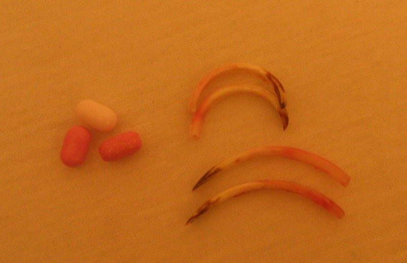
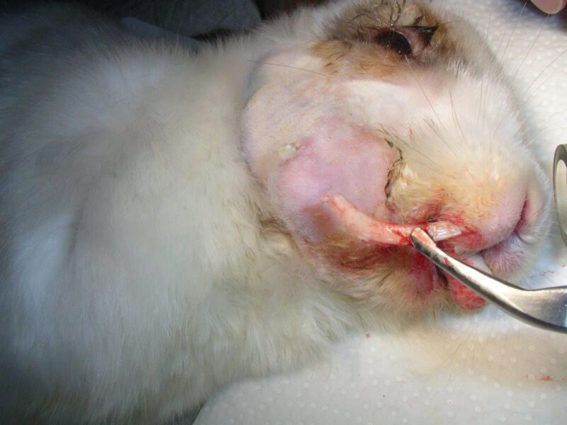
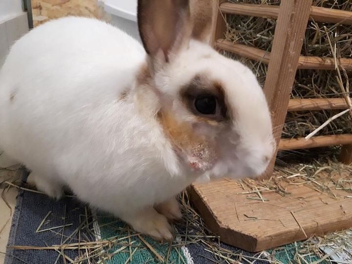
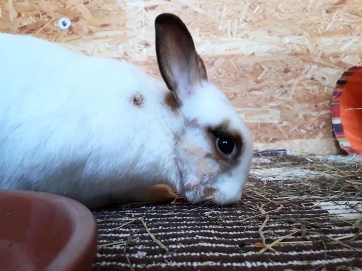
Case example: Jaw abscess caused by „tooth clipping“ – Before and After
Gizmo’s molars became severely infected as a result of „tooth clipping,“ which led to a jaw abscess. Despite intensive treatment, he didn’t survive and had to be euthanized. Gizmo was put to sleep at the age of four. This fate is shared by countless rabbits in Germany.
Common Mistake: Dental Treatment Without Anesthesia
“My vet treats my rabbit’s teeth without anesthesia.”
- This is something many rabbit owners hear — and believe.
However, this approach is outdated and potentially harmful.
Why “tooth clipping” is a problem
Many owners avoid anesthesia due to fear and choose tooth clipping using a mouth spreader instead.
👉 This is a serious mistake.
Clipping:
- does not treat the root cause
- does not correct the natural tooth alignment
- only temporarily reduces symptoms
As a result:
- teeth must be trimmed every 10–14 days
- the underlying disease continues to worsen
- the rabbit often suffers from ongoing pain
Why early proper treatment matters
By the time a rabbit is seen by a specialized dental veterinarian, the condition is often already advanced.
With early and correct treatment under sedation or anesthesia:
- the root cause can be identified
- proper tooth alignment can be restored
- natural chewing function can return
👉 In many cases, no further regular treatment is needed.
Real-life outcomes
Many rabbit owners report:
After months or even years of repeated tooth clipping,
a single proper dental correction by a specialist
→ eliminated the need for ongoing treatments.
Safety of sedation
Most dental procedures require only sedation, not full anesthesia.
The risks are generally low when performed correctly.
Well-treated rabbits can:
- live long lives
- tolerate repeated sedation if necessary
⚠️ Why clipping is dangerous
Clipping teeth in conscious rabbits can cause:
- splintering of the teeth
- microcracks extending to the roots
- infections and abscesses
In addition, stress and defensive movements can lead to:
- spinal injuries
- fractures
Important conclusion
– Tooth clipping without anesthesia is an outdated practice.
– Proper dental treatment focuses on:
- correct diagnosis
- gentle techniques
- long-term solutions
Case Reports
Moritz, a Satin Rabbit with Numerous Jaw Abscesses and Dental Problems
The entire lower right tooth row and one lower incisor were removed. Through surgeries and long-term antibiotics (Amoxicillin), the condition was brought under control, allowing him to live for several more years without abscesses.

Mia
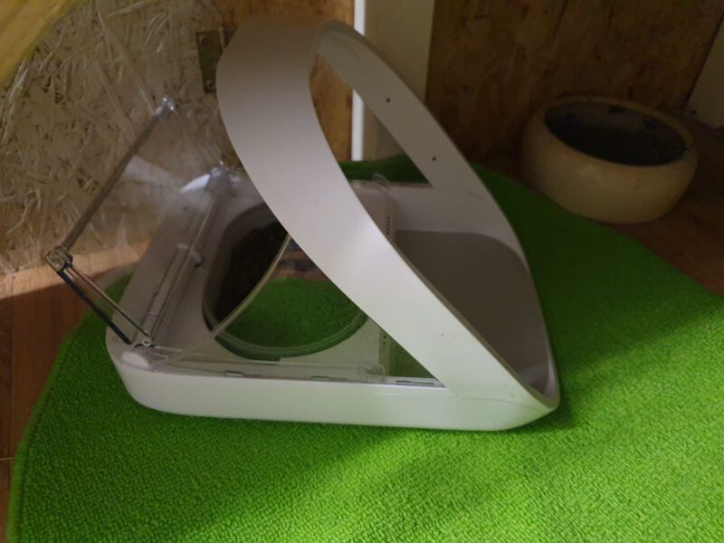
Mia had severe abscesses in two quadrants and overall very bad teeth. In several surgeries, all of her molars were removed (first the abscesses on the top right and bottom left, then gradually, depending on the severity). She still has all her incisors. One of her front teeth was affected at the root by the inflamed molars, but it grew back healthily after the diseased molars were removed. She now eats Cuni Complete from the automatic feeder (a handful twice daily) and vegetables (cucumber, cabbage, bell peppers… softer foods that she can bite into; no more grass). You wouldn’t even notice that she has no molars. Although I initially found the thought of a toothless rabbit strange, I would say I would do it again in a heartbeat. She got sick at 3 years old and is now 10 years old. After the dental treatment was completed, she hasn’t needed to see the vet for her teeth and has been completely healthy!

Nutrition for Dental Diseases
Nutritional imbalances are often the cause of dental diseases, so affected animals should be switched to a diet consisting solely of fresh leafy greens.
Improving tooth wear and potentially preventing the formation of tooth spurs, or extending the time between trims, can be read here: Tooth Wear.
Feeding After Dental Surgeries and for General Dental Diseases
However, if the incisors have been removed or if the animal has severe molar misalignments, a special diet is required.
Sources/Further Reading:
Böhmer, E., Crossley, D.(2009): Objective interpretation of dental disease in rabbits, guinea pigs and chinchillas. Use of anatomical reference lines. Schattauer
Böhmer, E. (2011). Zahnheilkunde bei Kaninchen und Nagern: Lehrbuch und Atlas; mit 27 Tabellen. Schattauer.
Böhmer, C., & Böhmer, E. (2017): Shape variation in the craniomandibular system and prevalence of dental problems in domestic rabbits: a case study in Evolutionary Veterinary Science. Veterinary sciences, 4(1), 5.
Böhmer, E. (2017): Frage an den Tierarzt/Tierärztin: „Sind die Zähne meines Kaninchens in Ordnung?“ [http://curoxray.de/blog/frage-an-den-tierarzttieraerztin-sind-die-zaehne-meines-kaninchens-in-ordnung/, 9.12.2017]
Ewringmann, A. (2016): Leitsymptome beim Kaninchen: diagnostischer Leitfaden und Therapie. Georg Thieme Verlag.
Ewringmann, A. (2017): Keimspektrum und Antibiotikasensitivitäten bei eitrigen Zahnerkrankungen von Kaninchen. Tierärztliche Praxis Ausgabe K: Kleintiere/Heimtiere, 45(06), 373-383.
Gabriel, S. (2013): Röntgendiagnostik bei Malokklusion des Kaninchens. veterinär spiegel, 23(01), 17-22.
Gabriel, S. (2014); Extraktion der Inzisivi bei Kaninchen. kleintier konkret, 17(S 01), 13-19.
Gabriel, S. (2016): Molarenextraktion bei Heimtieren–Indikation und Technik. kleintier konkret, 19(S 02), 18-22.
Gabriel, S. (2015): Praxisbuch Zahnmedizin beim Heimtier. Georg Thieme Verlag.
Glöckner, B. (2002): Untersuchungen zur Ätiologie und Behandlung von Zahn-und Kiefererkrankungen beim Heimtierkaninchen (Doctoral dissertation, Freie Universität Berlin, Universitätsbibliothek).
Glöckner, B. (2014): Spülung des Tränennasenkanals bei Dakryozystitis und Dakryostenose. kleintier konkret, 17(S 01), 27-30.
Glöckner, B. (2016): Dacryocystitis beim Kaninchen. team. konkret, 12(04), 8-13.
Gorrel, C. (2006). Zahnmedizin bei Klein-und Heimtieren. Elsevier, Urban&FischerVerlag.
Harcourt-Brown, F. M. (1996): Calcium deficiency, diet and dental disease in pet rabbits. The Veterinary Record, 139(23), 567-571.
Harcourt‐Brown, F. M., & Baker, S. J. (2001): Parathyroid hormone, haematological and biochemical parameters in relation to dental disease and husbandry in rabbits. Journal of Small Animal Practice, 42(3), 130-136.
Harcourt-Brown, F. (2006): Metabolic bone disease as a possible cause of dental disease in pet rabbits.
Harcourt-Brown, F. (2009): Dental disease in pet rabbits 1. Normal dentition, pathogenesis and aetiology. In Practice, 31(8), 370. [https://www.researchgate.net/profile/Frances_Harcourt-Brown/publication/298854593_Dental_disease_in_pet_rabbits_1_Normal_dentition_pathogenesis_and_aetiology/links/5794c62808ae33e89f97970b/Dental-disease-in-pet-rabbits-1-Normal-dentition-pathogenesis-and-aetiology.pdf, 9.12.2017]
Hartmann, M. (2007): Zahnextraktion hypsodonter Zähne bei Nagern und Hasenartigen. veterinär spiegel, 17(01), 22-27.
Jekl, V., & Redrobe, S. (2013): Rabbit dental disease and calcium metabolism–the science behind divided opinions. Journal of Small Animal Practice, 54(9), 481-490.
Korn, A. K. (2015): Zahn-und Kieferveränderungen beim Kaninchen: Diagnostik, Auftreten und Heritabilitäten.
Köstlinger, S. (2014): Vergleich der digitalen Röntgenuntersuchung mit der computertomographischen Untersuchung des Schädels bei zahnerkrankten Kaninchen(Doctoral dissertation, Bibliothek der Tierärztlichen Hochschule Hannover).