Contents
Inflammation/Discharge of the Nasolacrimal Duct (Dacryocystitis)
Eye Discharge in Rabbits
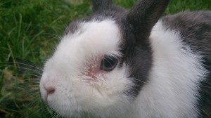
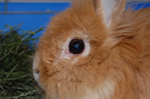
Dacryocystitis is an inflammation or blockage of the tear drainage system (lacrimal canaliculi, nasolacrimal duct, and/or lacrimal sac). This condition results in a watery or white, milky discharge from the eye, often leading to crusts in the fur and a wet area around the eye. If the fluid is clear, it is usually not caused by an infection. Some rabbits clean their partner’s eyes so thoroughly that this initial stage may go unnoticed. In later stages, thick, white pus oozes out. This pus can also originate from abscesses at the tooth roots that press into the nasolacrimal duct.
With this disease, other parts of the eye, such as the cornea, eyelids, and conjunctiva, are often inflamed as well, since the condition can spread.
What is the exact cause?
The causes of discharge or inflammation of the nasolacrimal duct in rabbits are almost always related to dental issues, often in combination with bacterial infections. In rabbits, the tooth roots are located very close to the nasolacrimal duct. When the teeth become too long (due to insufficient wear, retrograde root growth, or malocclusion of continuously growing teeth), the roots can compress or completely obstruct the nasolacrimal duct.
Inflamed tooth roots often lead to inflammation and pus in the nasolacrimal duct, or the inflammation can occur the other way around. More information can be found under „Jaw Abscesses.“
Rabbits with very round heads, such as lops and dwarf breeds, are genetically predisposed to dental diseases that affect the nasolacrimal duct (see Brachycephaly).
Retrograde tooth growth is mainly caused by improper nutrition (see below). Many rabbits are fed incorrectly, often without the owners realizing it! Over the years, this can lead to excessively long tooth roots and jaw abscesses.
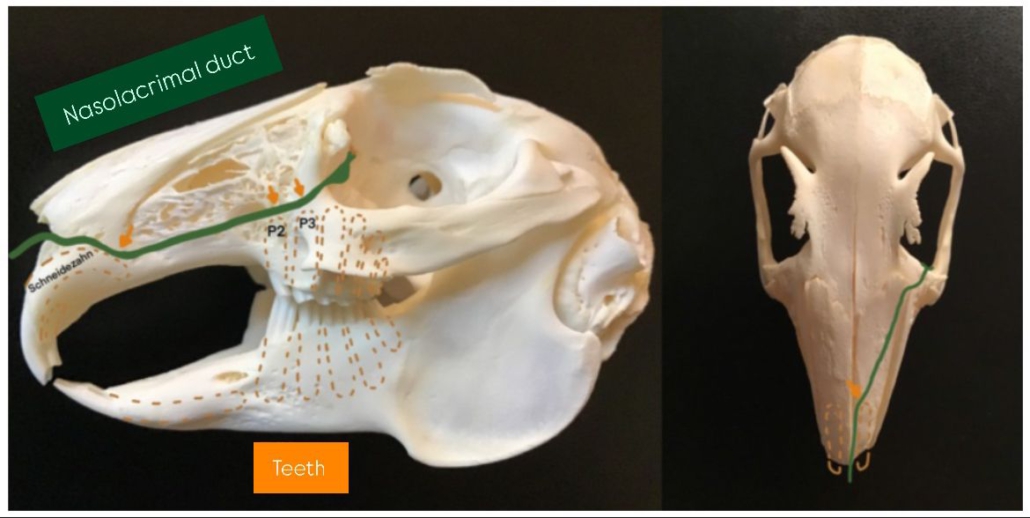
Brown: Non-visible part of the teeth.
The nasolacrimal duct runs very close to the tooth roots (orange arrows), so diseases of the upper jaw (retrograde tooth growth, jaw abscesses, etc.) often manifest only through nasal or eye discharge.
Why Do Rabbits Frequently Suffer from This Condition?
Rabbits Have a Unique Anatomy
It is often asked why rabbits, in particular, frequently suffer from watery eyes! Many veterinarians unfamiliar with rabbit-specific anatomy fail to diagnose and treat the condition correctly.
The nasolacrimal duct in rabbits runs from the lacrimal punctum, located in the inner corner of the lower eyelid (towards the nose), down to the nasal cavity. Unlike other domestic animals, such as horses, rabbits do not have a lacrimal punctum in the upper eyelid. Shortly after the lacrimal punctum, the nasolacrimal duct transitions into a lacrimal sac, where larger amounts of pus can accumulate if the duct becomes inflamed. The nasolacrimal duct then narrows again and passes very close to the root tips of teeth in two critical areas—especially near two premolars (P2 and sometimes P3), the foremost upper molars, as well as the roots of the incisors. The nasolacrimal duct opens laterally inside the nose.
These narrow passages near the tooth roots make the nasolacrimal duct highly susceptible to partial or complete blockage caused by even mild retrograde tooth growth (elongated tooth roots), leading to discharge.
Susceptibility to Infections
The accumulated tear fluid provides an ideal environment for bacterial growth, which often results in secondary infections and inflammation.
Constantly Growing, Open-Rooted Teeth
Rabbits have continuously growing, open-rooted teeth, meaning that feeding errors can have severe consequences. Improper diet leads to insufficient tooth wear or unnatural stress on the teeth, causing dental issues (see below).
Importance of Specialized Care
Since the unique anatomy and physiology of rabbits are not covered in standard veterinary education, it is crucial to consult a veterinarian who specializes in rabbit dentistry and care!
Weeping Rabbits?
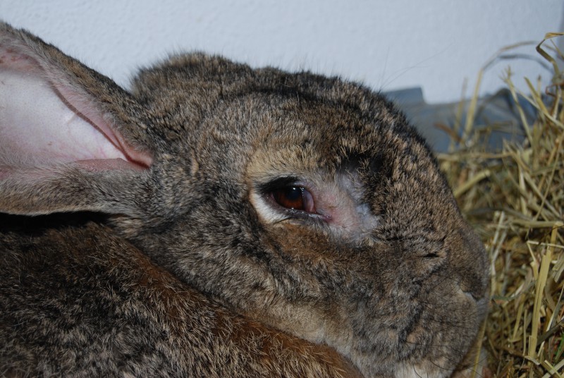
Often, singly housed rabbits experience clear discharge from their eyes, with no apparent cause, which disappears suddenly after they are paired with another rabbit. Some owners refer to this as their rabbit „crying.“ A possible explanation is that the partner rabbit, after bonding, cleans the discharge away, making it no longer visible.
In most cases, these rabbits also suffer from dental root diseases, but in the early stages, the eye care provided by the partner rabbit can mask the symptoms, making the condition less noticeable.
How the Veterinarian Proceeds…
- The eyes must be thoroughly examined. For example, it needs to be determined whether the rabbit has an eyelid inflammation, conjunctivitis (either as a cause or a consequence of the inflammation). It may also be necessary to stain the eye to rule out corneal injuries (keratitis), which often occur as a result of chronic inflammation.
- The Schirmer Tear Test 1 (STT1) is used to measure the amount of tear fluid in the eye. A special test strip is placed between the conjunctiva and cornea of the lower eyelid. The result is read from the scale of the test strip after 60 seconds. The height of the wetted strip indicates the amount of tear fluid produced (Reference values for rabbits: 5.3 mm/min ± 2.96, with a range between 0 and 15 mm/min or between 0 and 11.2 mm/min at double standard deviation (Abrams et al. 1990). There is an interaction effect between the eyes, as higher STT values are obtained when the second eye is tested. There are breed-specific differences: White New Zealand (7.58 ± 2.3 mm/min), NHD Lop 10.0 (5.0–17.3) mm/min).
- Breathing should be carefully checked, as inflamed nasolacrimal ducts can lead to respiratory infections or may even be the cause of them. The lungs and heart should also be auscultated, as they may also be affected.
- The oral cavity should be inspected. An otoscope, speculum, or cheek retractor is used for this, but not a mouth spreader attached to the front teeth. It must not be used on a conscious rabbit! The veterinarian will check for any foreign objects wedged between the teeth, or for loose or discolored teeth.
- The nasolacrimal duct will be flushed by the veterinarian first (but it is not always patent). If it is not patent, dental diseases are often the underlying cause. Tooth roots may grow into the nasolacrimal duct or compress it through swelling/inflammation. In some cases, the nasolacrimal duct may also be blocked or dilated due to chronic inflammation, or the secretions may clog it.
- It is important to determine whether the condition is related to dental problems (X-rays from at least four angles, see video), as this is almost always the cause. Retrograde growth (overly long tooth roots that penetrate the jaw) can only be detected through X-rays.
- By introducing contrast medium into the nasolacrimal duct and performing subsequent head X-rays (oblique views), the cause of the poor duct patency can be more precisely identified. CT scans (microCT) are superior to regular X-rays and may be used depending on the case.
- Please consult a rabbit dental specialist from this list if your rabbit has eye discharge. Other veterinarians may not be familiar with the specific anatomy of the nasolacrimal duct and its connection to dental diseases.
Treatment
Addressing the Cause
The underlying cause of the eye discharge must be identified and treated (e.g., dental treatments); otherwise, the discharge will continue to reappear. In the case of retrograde tooth growth, the teeth are corrected under anesthesia and filed down to a length that prevents the rabbit from chewing on them for a short period. This helps relieve pressure on the nasolacrimal duct and stops the retrograde growth. In some cases, affected teeth may need to be extracted, especially if there are abscesses on the tooth roots, which might require the removal of the infected teeth (see jaw abscesses). X-rays are used to assess and rule out other conditions. Proper dental correction will eventually result in the disappearance of the eye discharge.
A less common cause can be infectious diseases (e.g., myxomatosis or rabbit snuffles), conjunctivitis spreading to the nasolacrimal duct, or underlying allergic conditions. If not treated promptly, these can also cause the nasolacrimal duct to become enlarged, inflamed, and blocked.
Make sure to see a rabbit-savvy veterinary dentist, as regular veterinarians do not have sufficient dental skills. Or do you go to your general doctor when you have tooth pain?
After the dental correction, appropriate aftercare is important, including the right pain medication, supplemental feeding, and intensive care for the animal. It is best to schedule the correction so that you can take care of the rabbit intensively yourself, especially on the day of the procedure and for the two to five days following the correction.
Treatment without dental correction?
We frequently encounter cases where rabbits are only treated with antibiotic eye drops without identifying and treating the affected teeth. Often, even dental X-rays in multiple planes have not been performed. In these cases, the inflammation may subside with the drops, but it returns after a short or sometimes longer period. Often, a whole list of different drops and ointments is prescribed, instead of addressing the underlying cause. Such treatment will never be successful if the cause remains! We often hear of cases where the eye is simply left untreated in the long term, or only the surrounding area is regularly washed, since the treatment „doesn’t work anyway.“
Such an approach is not in line with animal welfare! The rabbit suffers from the inflammation, experiences pain, and has to live with secondary conditions, such as eye infections, an enlarged nasolacrimal duct due to chronic inflammation, the development of (further) tooth root abscesses from the inflammation near the tooth roots, pain from retrograde tooth roots, and even pressure on the eye, etc.! Many of these conditions are not visible externally, but the animals often show signs of pain.
Matting in Fur
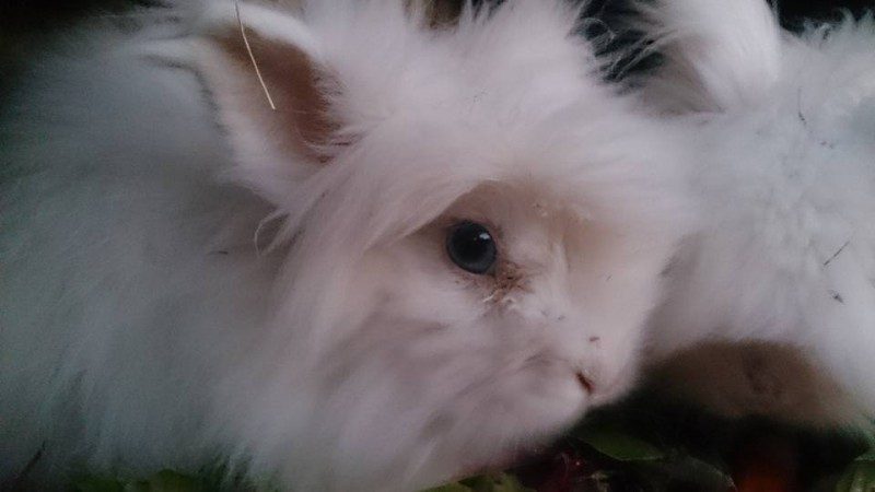
It is often not easy to remove the matting in the fur. You can try gently cleaning and softening the eye area with a damp cloth, and then combing out the secretion with a flea comb.
Adjustment of Housing: Relieving the Airways and Reducing Matting
During housing, it is important to ensure good ventilation, frequent bedding changes (to avoid ammonia buildup), and use of dust-free bedding to relieve the respiratory system.
Additionally, it may be necessary to temporarily limit digging opportunities. This can help reduce matting in the fur.
Are Watery Eyes Really Only Due to Allergies?
It is only after dental X-rays by a rabbit-savvy veterinary dentist and thorough flushing of the nasolacrimal duct (multiple times) that this diagnosis is made! If allergies are truly the cause, eye drops for care can be used as a long-term treatment. Sometimes, the trigger can be addressed, such as changing the bedding. However, this is very rare. Most rabbits diagnosed with „allergies“ actually have dental problems.
Nutrition is Key!
Watery eyes are often diet-related. Years of improper nutrition can lead to misalignment of the teeth, causing the rabbit to chew unnaturally.
Rabbits chew leafy fresh foods (such as green forage) with their teeth using grinding movements (sliding the teeth back and forth). Inappropriate foods like dry food, pellets, extrudates, carob, and pea flakes are crushed by „biting together.“ This creates an unnatural pressure on the tooth roots, pushing the teeth further into the jaw. In extreme cases, the teeth can compress or even perforate the nasolacrimal duct, which runs close to some of the teeth. This blockage prevents the tear fluid from draining, causing watery eyes. The blockage in the nasolacrimal duct can also lead to inflammation, often affecting the tooth roots as well—resulting in pus discharge and jaw abscesses.
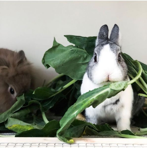
Healthy rabbits should be fed at least 80% fresh green food, with the remainder consisting of hay, small amounts of firm vegetables (such as carrots, celery, parsnips), and occasional small fruit treats from hand feeding.
If a rabbit is already suffering significantly from retrograde tooth growth, it is crucial to feed it an optimal diet. In summer, it should be fed solely with meadow herbs, and in winter with leafy vegetables (bitter greens, leafy cabbage varieties, vegetable tops, leafy vegetables…). Anything that adds additional pressure on the tooth roots must be completely eliminated from the diet. However, small amounts of carrot as a hand-fed treat can still be given. Dry food, seeds, oats, commercial treats, and pea flakes must be completely removed from their diet!
Sources include:
Abrams KL, Brooks DE, Funk RS, Theran P. (1990): Evaluation of the Schirmer tear test in clinically normal rabbits. Am J Vet Res 1990; 151: 1912–3
Biricik HS, Oguz H, Sindak N, Gurkan T, Hayat A. (2005): Evaluation of the Schirmer and phenol red thread tests for measuring
tear secretion in rabbits. Vet Rec 156:485–487.
Böhmer, E. (2011): Zahnheilkunde bei Kaninchen und Nagern: Lehrbuch und Atlas; mit 27 Tabellen. Schattauer.
Corsi, F., Arteaga, K., Corsi, F., Masi, M., Cattaneo, A., Selleri, P., … & Guandalini, A. (2022): Clinical parameters obtained during tear film examination in domestic rabbits. BMC Veterinary Research, 18(1), 398.
Ewringmann, A. (2016): Leitsymptome beim Kaninchen: diagnostischer Leitfaden und Therapie. Georg Thieme Verlag.
Gabriel, S. (2013): Röntgendiagnostik bei Malokklusion des Kaninchens. veterinär spiegel, 23(01), 17-22.
Gabriel S. (2016): Praxisbuch Zahnmedizin beim Heimtier. Stuttgart: Enke
Glöckner, B. (2002): Untersuchungen zur Ätiologie und Behandlung von Zahn-und Kiefererkrankungen beim Heimtierkaninchen, Diss.
Glöckner, B. (2014). Spülung des Tränennasenkanals bei Dakryozystitis und Dakryostenose. kleintier konkret, 17(S 01), 27-30.
Glöckner, B. (2016): Dacryocystitis beim Kaninchen. team. konkret, 12(04), 8-13.
Whittaker, A. L., & Williams, D. L. (2015): Evaluation of lacrimation characteristics in clinically normal New Zealand white rabbits by using the Schirmer tear test I. Journal of the American Association for Laboratory Animal Science, 54(6), 783-787.
Walde I, Nell B, Schäffer E, Köstlin R, Hrsg. (2008): Augenheilkunde. 3., überarbeitete und erweiterte Auflage. Stuttgart: Schattauer


