
Otitis (Ear Infections)
Unfortunately, ear infections are relatively common in rabbits. Studies of adult rabbits from commercial meat-production systems have found signs of otitis in around 32% of animals. Depending on the breed, this is broadly consistent with observations reported in German veterinary practices.
Risk factors for ear infections in rabbits
Several factors can increase the risk of ear infections in rabbits, including breed, ear anatomy, mobility problems and respiratory disease.
- Lop-eared rabbits are particularly prone to ear infections and otitis media. An infection of the outer ear can spread to the middle ear. Many lop-eared rabbits develop ear problems during their lifetime. Their ear anatomy differs from that of upright-eared rabbits: the cartilage fold that causes the ears to droop creates a bend in the ear canal, which can make the natural removal of earwax more difficult. Studies have also found that the ear canals of lop-eared rabbits are significantly narrower than those of upright-eared rabbits. In addition, the hanging ear flap reduces ventilation of the ear canal. Together, these factors can create conditions that promote inflammation.
- Very small ears, as seen in some dwarf rabbit breeds, may also increase the risk because the ear canal can be particularly narrow.
- Heavily furred ears may interfere with ventilation and the natural removal of earwax, which can also contribute to ear problems.
- Very large rabbit breeds appear to be affected more frequently as well. One possible explanation is their increased tendency to develop joint disease. Rabbits with reduced mobility may have difficulty grooming and cleaning their ears properly. Large rabbits may also come more frequently from populations affected by ear mites, which can trigger inflammation of the ear canal.
- Rabbits with joint problems such as osteoarthritis or spondylosis, an amputated leg or other mobility disorders may be at increased risk because they can no longer clean their ears effectively on their own.
- Neurological disorders may also be associated with ear disease. In one study, around 63% of rabbits with neurological symptoms, such as seizures or head tilt, were also found to have ear infections.
- The reported proportion increased to as much as 85% when a respiratory disease was also present. Bacteria and other pathogens from the nasal passages can spread to the middle ear. In another study, up to 30% of rabbits with respiratory disease associated with pasteurellosis had otitis media despite showing no obvious external signs.
Symptoms of ear infections in rabbits: how to recognise them
Ear infections in rabbits can be surprisingly difficult to detect. Some rabbits show very few obvious signs, and in one study almost one in three rabbits with an ear infection showed no clinical symptoms despite a thorough examination. This is one reason why otitis can remain unnoticed for a long time.
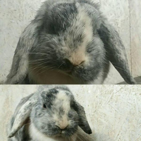
Some affected rabbits simply become quieter or less active because of pain. In rarer cases, they may become irritable or unusually aggressive. Lop-eared rabbits in particular may have heavily soiled ear canals, and in some cases the canal can become partly or completely blocked by earwax and debris.
Possible signs of an ear infection include:
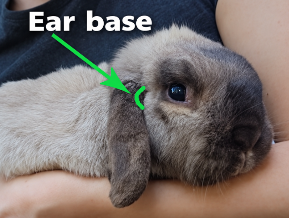
- Small lumps or swellings at the base of the ear, which may indicate an ear-base abscess.
- Ear bases that appear red or feel unusually warm or cold.
- Pain when the base of the ear is carefully touched or when the rabbit is stroked around the head.
- Head shaking or scratching at the ears, particularly in some infections involving yeast or irritation of the ear canal.
- Repetitive swaying or scanning movements of the head.
- Nystagmus, meaning rapid or involuntary eye movements.
- An asymmetrical or crooked appearance of the mouth or face.
- Reduced hearing or deafness.
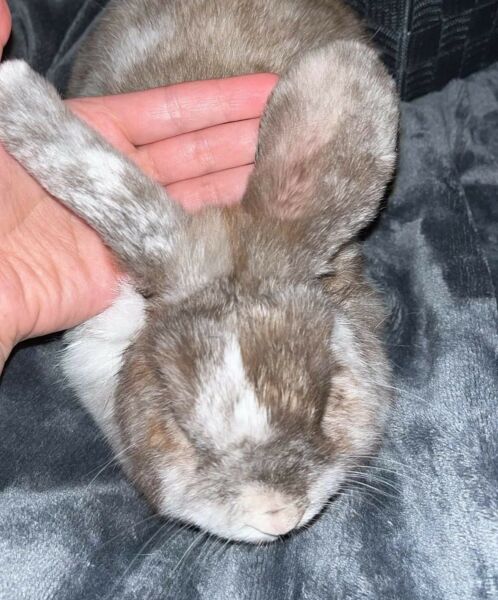
When checking the base of the ears, it can be helpful to gently compare both sides at the same time. Differences in shape, firmness or swelling may make an abscess or other abnormality easier to notice.
Rabbits that have become hard of hearing or deaf may startle when approached from outside their field of vision. Owners sometimes mistake this behaviour for very deep sleep, because the rabbit no longer reacts normally to sounds.
Aural haematoma
An aural haematoma can occasionally develop in connection with an ear infection, although this appears to be relatively uncommon in rabbits.
Caption: Aural haematoma associated with an ear infection in a rabbit.
Swelling at the base of the ear
Changes or pouch-like swellings around the ear canal may sometimes be detected by carefully feeling and comparing the base of both ears.
Caption: Swelling at the base of the ear can sometimes be detected by gently comparing both sides.
Advanced ear infections and neurological symptoms
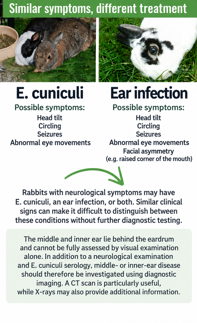
Advanced ear infections in rabbits, especially when the middle or inner ear is affected, can cause neurological signs that may resemble E. cuniculi. For this reason, the two conditions can sometimes be confused.
Possible neurological signs include:
- Head tilt
- Loss of balance or unsteady movement
- Walking or circling repeatedly in one direction
- Nystagmus or abnormal eye movements
- Problems with coordination
Because these symptoms can have several different causes, a rabbit showing head tilt, balance problems, circling or abnormal eye movements should be examined promptly by a rabbit-experienced veterinarian.
Possible complications of chronic ear infections in rabbits
Chronic inflammation and persistent pain can place considerable strain on an affected rabbit. Rabbits with long-standing ear disease may also develop or experience other health problems, including E. cuniculi, parasitic infections, tooth-root inflammation, abscesses and respiratory disease.
Chronic pain in the head and ear area can also change the way a rabbit chews. Over time, altered chewing patterns may contribute to dental disease, including problems involving the tooth roots. Dental problems are therefore frequently seen alongside chronic ear disease.
Some rabbits eat less because of pain. In more severe cases, they may stop eating altogether. Reduced food intake can quickly lead to gastrointestinal problems, making loss of appetite an important warning sign in rabbits.
Further information on specific symptoms
- Nystagmus (abnormal eye movements): Horizontal or rotational involuntary eye movements are particularly common.
- Aggressive or unusual behaviour: A small proportion of rabbits with chronic ear pain may become more irritable, less tolerant of other rabbits or more difficult to bond. Behavioural changes can be a sign of underlying pain.
- Facial asymmetry or a crooked mouth: Unilateral facial nerve paralysis or facial nerve spasms may occur. Initially, facial nerve paralysis may cause the corner of the mouth to droop on the affected side. In chronic cases, a facial nerve spasm may instead pull the corner of the mouth upward on the affected side (ipsilateral hemifacial spasm), making the opposite side appear to droop. The affected eye may also remain partly closed, the third eyelid may protrude, or normal eyelid closure may be impaired. Signs consistent with Horner’s syndrome may also occur.
- Balance problems: These can occur with an inner ear infection (otitis interna) when the vestibulocochlear nerve is affected. This nerve is involved in both balance and hearing.
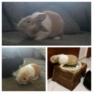
Leo developed head tilt, seizures, respiratory symptoms and left-sided weakness/paralysis. For around six months, he was treated for suspected E. cuniculi, including courses of three different antibiotics. Only later, during a consultation, was his owner advised that an ear infection could be responsible for the neurological symptoms. Further examination confirmed an ear infection.
By the time the correct diagnosis was made, Leo’s condition had already deteriorated significantly and he had become markedly stiff.
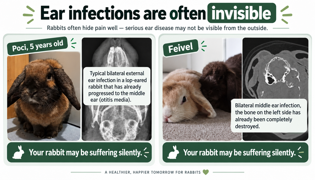
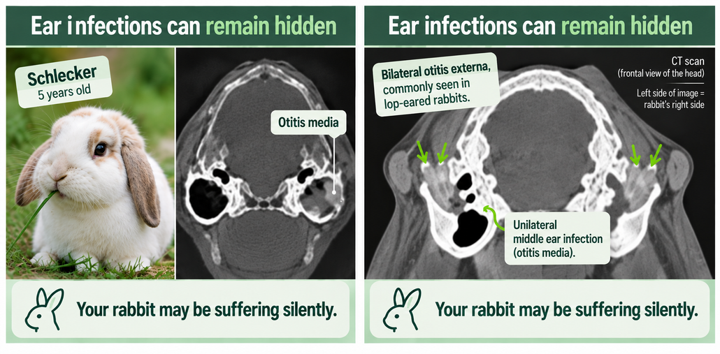
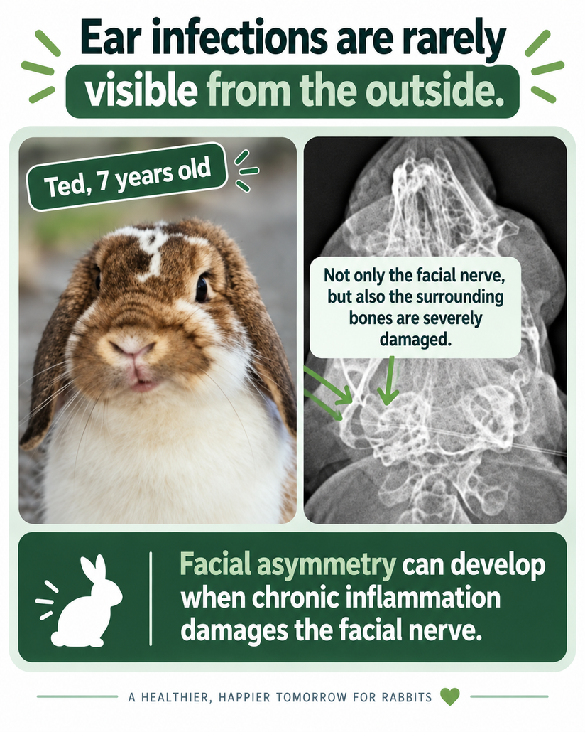
| Type of ear infection | Area affected | Possible signs | How it is diagnosed |
|---|---|---|---|
| Otitis externa | External ear canal | Earwax or discharge, scratching, head shaking, pain, swelling at the base of the ear | Otoscopic or endoscopic examination, cytology, bacterial culture if required |
| Otitis media | Middle ear behind the eardrum | May cause few obvious signs; pain, reduced activity, facial asymmetry, hearing problems or neurological signs may occur | CT or CBCT is particularly useful; X-rays may detect more advanced disease |
| Otitis interna | Inner ear | Head tilt, balance problems, circling, nystagmus and other neurological signs | CT or MRI; MRI may be particularly useful when neurological or soft-tissue involvement is suspected |
Diagnosis of ear infections in rabbits
Find a veterinarian experienced in rabbit ear disease
If you suspect an ear infection, it is essential to consult a veterinarian who is experienced not only with dogs and cats, but specifically with rabbits and other small mammals.
Rabbit medicine receives comparatively limited attention during veterinary training. Diagnosing and treating ear infections in rabbits therefore requires additional training, experience and appropriate diagnostic equipment.
Why ear infections in rabbits are easily missed
Many rabbits with an ear infection show no obvious outward signs. Sometimes the only clues are slightly dirtier ear canals, reduced activity or unusually quiet behaviour.
Ear disease may occasionally be discovered while investigating abnormal blood results. Elevated inflammatory parameters, such as leukocytosis or a pseudo-left shift, may prompt the veterinarian to look for an underlying source of inflammation. However, not every rabbit with an ear infection develops detectable inflammatory changes in the blood. Normal blood results therefore do not rule out otitis.
Several parts of the ear may be affected at the same time.
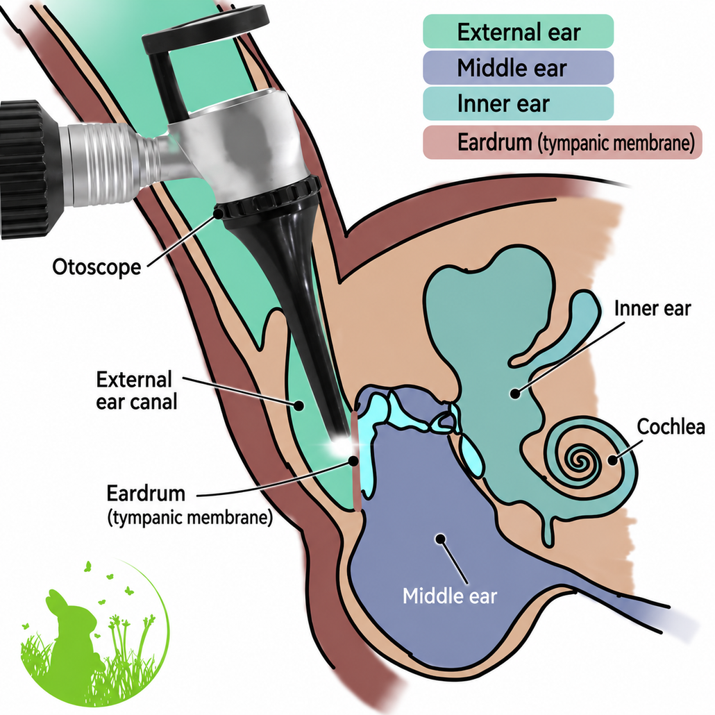
With an otoscope, the veterinarian can examine only the external ear canal. Inflammation behind the eardrum, including middle and inner ear infections, cannot be seen directly.
Otitis externa – infection of the external ear canal
Otitis externa affects the external ear canal and may sometimes be detected by examining the ear with an otoscope or endoscope.
However, the inflammation can easily be hidden by:
- accumulated earwax
- narrowing of the ear canal
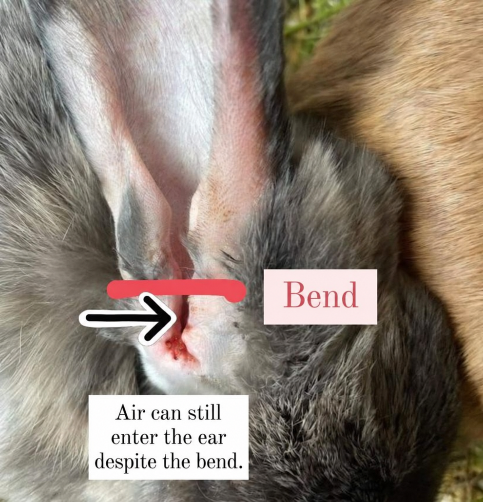
- the characteristic bend in the ear canal of lop-eared rabbits
In some lop-eared rabbits, gently lifting the ear and using suitable equipment allows an experienced veterinarian to examine the area beyond this bend. This is important because inflammation is often located deeper in the ear canal and may otherwise be missed.
A sample taken from deeper within the ear canal may help to distinguish inflammatory or purulent material from ordinary earwax. Normal rabbit earwax is usually pale yellow or beige, whereas pus is often paler and creamier, although the two can sometimes be difficult to distinguish by appearance alone.
Further testing may include:
- examination for ear mites
- cytology to assess cells and microorganisms under the microscope
- bacterial culture
- antibiotic susceptibility testing when appropriate
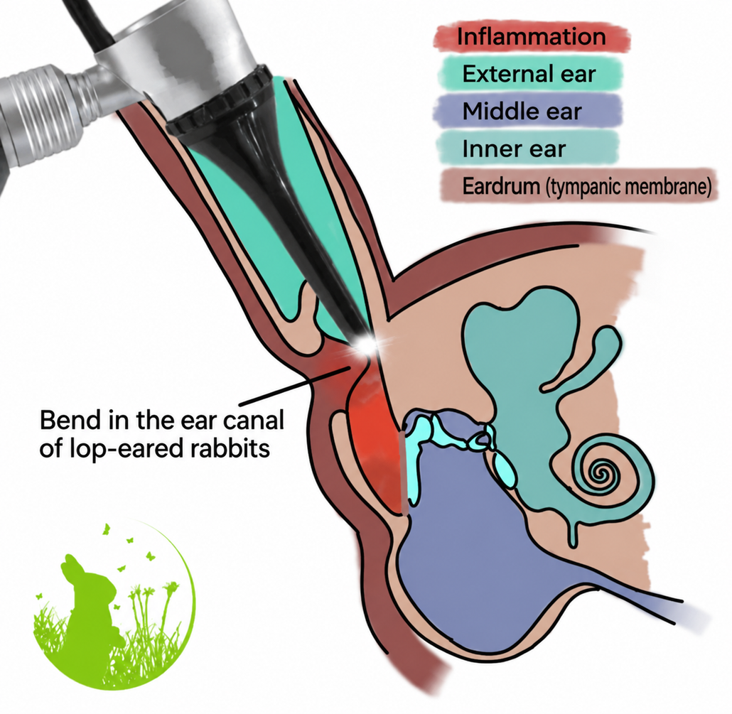
In lop-eared rabbits, the lower part of the ear canal is often difficult or impossible to visualise. Veterinarians who are not experienced in rabbit medicine may therefore consider the ears “clean”, even though inflammation is present beyond the bend in the ear canal.
Otitis media – middle ear infection
The middle ear lies behind the eardrum, which means that otitis media cannot normally be diagnosed simply by looking into the ear canal.
In severe cases, the eardrum may bulge outward or pus may enter the external ear canal. More commonly, however, middle ear infections remain completely hidden from view.
In lop-eared rabbits, examining the eardrum can be especially difficult because the ear canal is often narrow, curved and obstructed by earwax or inflammatory material.
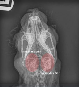
Standard X-rays usually reveal only more advanced middle ear disease, particularly once the surrounding bone has already been affected. A dorsoventral skull X-ray may provide useful information, but early changes can easily be missed.
More sensitive imaging techniques include:
- CT
- cone-beam CT (CBCT)
- MRI
These techniques are considerably more useful for detecting early or hidden middle ear disease and assessing the extent of the infection.
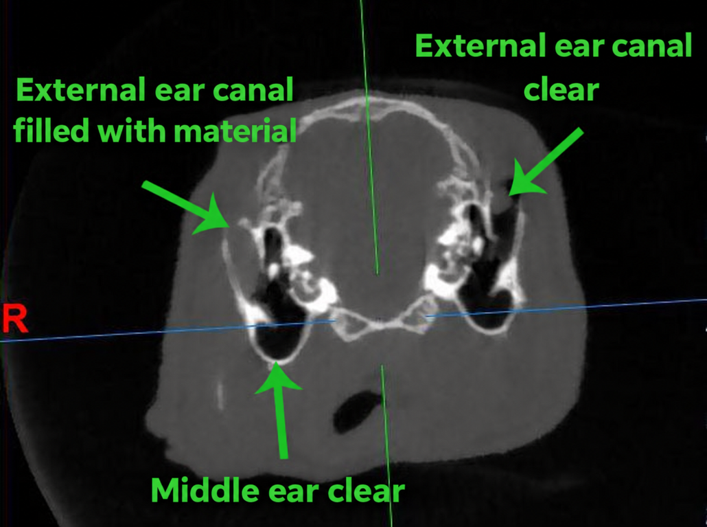
Image kindly provided by veterinarian Claus Meyer.
Otitis interna – inner ear infection
Otitis interna, or an inner ear infection, can be particularly difficult to diagnose using conventional X-rays, even when several views are taken.
Advanced imaging is often required. Depending on the individual case, CT, CBCT or MRI may be needed to assess the inner ear and surrounding structures.
MRI can be particularly useful when soft tissues, nerves or possible involvement of structures within the skull need to be evaluated.
Ear infections are often diagnosed too late
Unfortunately, many rabbits with chronic ear disease go through a long period of treatment before the underlying problem is correctly identified.
A particularly important example is head tilt in rabbits.
In one study, around one in three rabbits presenting with head tilt was found to have an ear infection rather than E. cuniculi. Both conditions can cause very similar neurological symptoms, and in some rabbits they may occur at the same time.
Owners sometimes report that their rabbit has been treated for presumed E. cuniculi for months without improvement, even though no diagnostic investigation has been performed to confirm that E. cuniculi is actually responsible for the symptoms.
A positive E. cuniculi antibody titre does not by itself prove that E. cuniculi is causing the rabbit’s current neurological signs. Many rabbits have been exposed to the organism without having active disease. Serology must therefore be interpreted together with the clinical findings and other diagnostic results.
When a rabbit develops neurological symptoms such as:
- head tilt
- loss of balance
- circling
- nystagmus
- facial asymmetry
- weakness or paralysis
middle and inner ear disease should always be considered and appropriately investigated.
In some cases, even the external ear has not been examined thoroughly before treatment for presumed E. cuniculi is started.
What can happen if an ear infection remains untreated?
If an ear infection remains undiagnosed for a long time, it may progress and become considerably more difficult to treat. The infection can also spread beyond the ear.
Possible complications of advanced or untreated ear disease include:
- dental and tooth-root disease
- abscesses
- meningitis
- inflammation involving the spinal cord or surrounding tissues
- spread of infection to the lungs or other organs
- involvement of joints
- severe destruction of the surrounding bone
These secondary problems can cause additional neurological or systemic symptoms, which may make the condition even more difficult to distinguish from E. cuniculi.
In severe chronic cases, the surrounding bone may become extensively damaged or destroyed. If the disease has progressed too far for effective treatment and the rabbit’s quality of life cannot be restored, euthanasia may ultimately have to be considered.
Because rabbits often show very few obvious signs of pain, they may live with significant pain for a long time without it being recognised.
E. cuniculi may become clinically active more readily in rabbits that are already weakened or under physiological stress. Some rabbits with a severe or chronic ear infection may therefore develop signs of E. cuniculi at the same time.
For this reason, even when an E. cuniculi antibody titre is positive or high, the ears should still be examined carefully. A positive titre alone does not prove that E. cuniculi is responsible for the rabbit’s current neurological symptoms, and middle or inner ear disease should not be overlooked.
Awake CT examination
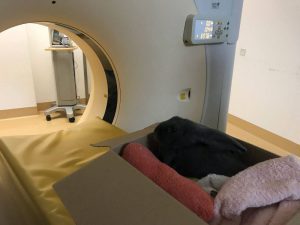
At the University of Veterinary Medicine Hannover, CT scans can in selected cases be performed while the rabbit is awake. The rabbit is placed in a suitable box, which is closed for the few minutes needed to complete the scan. The box is adequately ventilated so that the rabbit receives sufficient air throughout the procedure.
This can provide a gentle, minimally stressful form of diagnostic imaging and is increasingly used by veterinary centres where suitable equipment and protocols are available.
To investigate suspected ear infections in rabbits, diagnostic imaging of the head is often required. Skull X-rays can provide useful information, but they may fail to detect some cases of otitis media, particularly at an early stage.
For this reason, computed tomography (CT) is generally more suitable for assessing the middle ear and surrounding structures and can reveal changes that are not visible on conventional X-rays.
What causes ear infections in rabbits?
A middle ear infection (otitis media) can develop in two main ways. Infection may spread from an external ear infection (otitis externa) through a damaged or perforated eardrum into the middle ear. Alternatively, bacteria may spread from the nasal cavity through the auditory (Eustachian) tube, particularly in rabbits with respiratory infections or chronic nasal discharge.
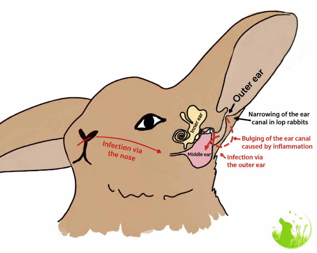
In lop-eared rabbits, part of the ear canal is often narrowed or obstructed by a characteristic bend. This can impair the normal removal of earwax and alter the environment within the ear canal, increasing the risk of inflammation.
Over time, the ear canal may develop an outpouching, and an abscess can form at the base of the ear.
An infection of the external ear can spread into the middle ear, particularly if the eardrum becomes damaged or perforated. Infection can also spread from the nasal cavity to the middle ear via the auditory (Eustachian) tube, particularly in rabbits with respiratory disease.
Possible causes and risk factors for ear infections in rabbits include:
- Bacterial infections: Both Gram-positive and Gram-negative bacteria may be involved. In one reported series of bacterial cultures, approximately 80% of infections involved a single bacterial species, while just over 20% involved more than one species. Frequently identified bacterial families included Staphylococcaceae (45%), Enterobacteriaceae (26.1%), Pasteurellaceae (11.5%), Pseudomonadaceae (6.8%) and Streptococcaceae (4.3%).
- Ear mites (Psoroptes cuniculi) and the inflammation they cause.
- Bite wounds and abscesses around the ear.
- Dental disease and dental abscesses, which may contribute to infection in nearby structures.
- Poor ventilation and impaired earwax clearance, particularly in lop-eared rabbits, rabbits with very small or heavily furred ears, and rabbits with naturally narrow ear canals.
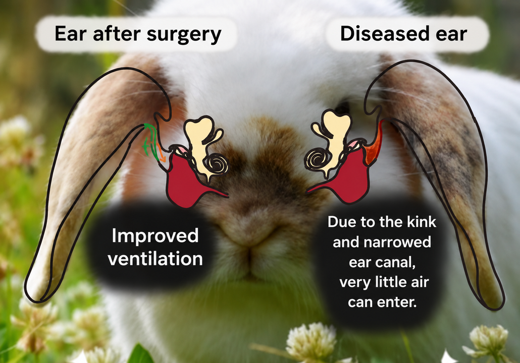
In lop-eared rabbits, the ear canal is anatomically narrower and develops a characteristic bend. This can be compared to a kinked garden hose: ventilation is reduced and earwax cannot be removed as efficiently. These anatomical changes can create conditions that promote chronic inflammation and ear infections.
Treatment of ear infections in rabbits
A rabbit with a suspected ear infection should be examined and treated by a veterinarian who is experienced in rabbit medicine and ear disease. Treatment depends on which part of the ear is affected, how advanced the disease is, whether the eardrum is intact, and which microorganisms or parasites are involved.
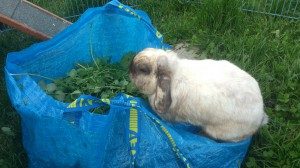
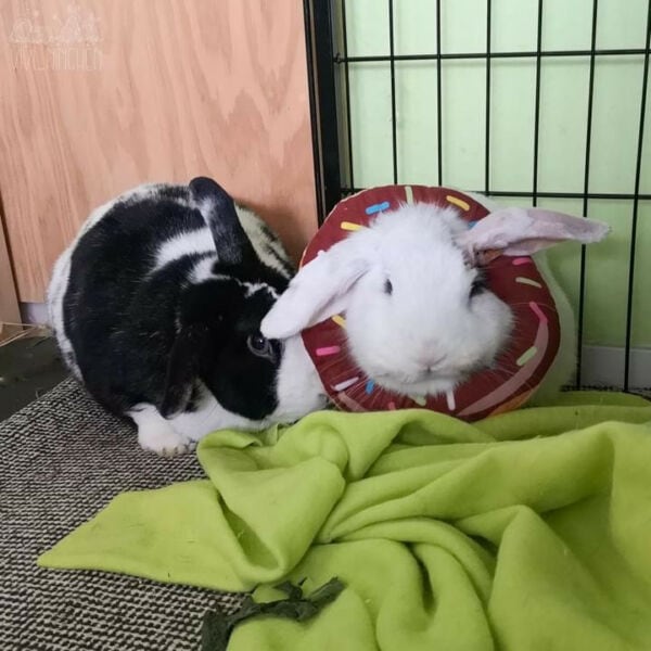
Ernie repeatedly injured other rabbits, sometimes severely, and eventually had to be re-bonded with new companions. Despite being seen by veterinarians experienced in rabbit medicine, no one initially considered taking skull X-rays or performing ear cytology.
His chronic ear infection was only diagnosed much later. By that time, the long-standing illness had already affected his social behaviour: he had dropped in the group hierarchy and had become noticeably more submissive.
Aggressive or unusually irritable behaviour in rabbits can be a sign of pain. In addition to reproductive disorders such as uterine disease, pain originating in the head—particularly from the ears or teeth—should always be considered as a possible underlying cause.
Treatment of otitis externa
Treatment of an external ear infection (otitis externa) should be based on the underlying cause rather than using the same medication for every rabbit.
Identifying the cause
Many different microorganisms can cause or contribute to otitis externa in rabbits. Cytology is particularly useful: a sample from the ear canal is stained and examined under a microscope to identify bacteria, yeasts, inflammatory cells or other abnormalities.
In some cases, bacterial culture and antibiotic susceptibility testing may also be recommended. This can help determine which bacteria are present and which antibiotic is most likely to be effective.
Samples should ideally be collected from the affected deeper part of the ear canal by the veterinarian, rather than simply from superficial earwax or debris.
Bacterial ear infections
If bacteria are involved, the veterinarian may prescribe a suitable topical antibiotic preparation. In some cases, an ophthalmic antibiotic preparation may be used off-label when an appropriate veterinary ear preparation is not available.
Treatment may be adjusted according to the results of cytology, bacterial culture or antibiotic susceptibility testing.
Particular care is required when the eardrum may be damaged or perforated, because some substances can be ototoxic and may damage structures of the middle or inner ear.
Enrofloxacin-containing preparations are sometimes used in rabbits under veterinary supervision. Depending on the individual case, compatible ear cleansers such as Tris-EDTA-based solutions may also be used to modify the environment within the ear canal and support antimicrobial treatment.
Topical medication can achieve very high concentrations directly within the ear canal. However, rabbits with middle ear disease, deeper infection or severe otitis may also require systemic antibiotics. Whether systemic treatment is necessary depends on the individual rabbit and the extent of the disease.
Yeast infections
If yeasts are identified, the veterinarian may prescribe an appropriate topical antifungal treatment.
Specially prepared miconazole solutions may be used in selected cases. The choice of preparation is important because some commercially available products contain ingredients that may be unsuitable, particularly if the eardrum is not intact.
In some cases, a systemic antifungal medication, such as itraconazole, may also be considered by the treating veterinarian.
Ear mites
If ear mites (Psoroptes cuniculi) are responsible for the inflammation, they must be treated with an appropriate antiparasitic medication.
Veterinary medicines containing substances such as selamectin or ivermectin may be used according to the veterinarian’s treatment plan.
Cleaning the external ear canal
Thorough cleaning is an important part of treating many ear infections in rabbits, because accumulated earwax, pus and debris can prevent medication from reaching the affected tissue.
In rabbits with severely narrowed or blocked ear canals, the first thorough cleaning may need to be performed under sedation or anaesthesia.
In lop-eared rabbits, particular attention must be paid to the area beyond the characteristic bend in the ear canal. Secretions frequently accumulate close to the eardrum behind this bend and may be difficult or impossible to see during a routine examination.
Appropriate flushing equipment may therefore be required to reach and clean the deeper part of the ear canal safely.
Ceruminolytic ear cleansers can help soften and remove excessive earwax. Thick purulent material may require more extensive flushing. In selected cases, the veterinarian may also use a mucolytic agent such as acetylcysteine (NAC) to help break down thick secretions.
Long-term ear care
Once the ear canal has been thoroughly cleared, regular cleaning may be necessary to reduce the risk of recurrent accumulation of earwax, microorganisms and inflammatory material.
The choice of ear cleanser and how frequently it should be used depend on:
- the type of ear infection
- the microorganisms present
- the amount of earwax produced
- the anatomy of the ear canal
- the severity of the disease
- the rabbit’s response to treatment
The veterinarian should determine the most appropriate cleaning schedule for the individual rabbit.
Some lop-eared rabbits with chronically narrow ear canals require long-term or lifelong ear care because their anatomy continues to impair ventilation and the normal removal of earwax.
In severe or repeatedly recurring cases, surgical widening or restructuring of the ear canal may be considered.
Ear cleansers can modify the environment within the ear canal and may help reduce bacterial or yeast overgrowth. The most suitable product should therefore be selected according to the findings in the individual ear rather than chosen at random.
If a rabbit develops redness, skin irritation, marked discomfort or signs of burning after an ear cleanser is used, the product should not simply be continued. The veterinarian should be contacted so that the ear can be reassessed and a more suitable preparation selected.
Important: ear medication must be chosen carefully
Not every ear preparation is suitable for rabbits, and some products may be unsafe when the eardrum is damaged or perforated.
Products containing antibiotics, antifungals, corticosteroids or other active ingredients can differ considerably in their effects and potential risks. Ear medication should therefore be selected specifically for the individual rabbit by a veterinarian experienced in rabbit medicine.
Important: Regular flushing and cleaning of the ear canal, together with an appropriate ear cleanser suited to the individual condition of the ear canal, are crucial to successful treatment. If treatment is not working as expected, common reasons include insufficiently frequent flushing, an unsuitable cleanser or incorrect application.
Initially, the ear should be thoroughly cleaned and flushed with a ceruminolytic ear cleanser to soften and remove accumulated earwax. This may need to be repeated until the ear canal is clear. Hypochlorous acid may then be applied to the ear canal. At home, a Tris-EDTA-containing ear cleanser may be used once or twice daily to modify the environment within the ear canal and help disrupt bacterial biofilms, which may improve the effectiveness of antibiotic treatment. Antimicrobial medication can usually be applied approximately 15 minutes later, depending on the products prescribed by the veterinarian.
Treatment generally continues for around six to eight weeks. In lop-eared rabbits, if the ear canal has not been surgically widened, ongoing ear care may be required for life to reduce the risk of recurrence. Depending on the individual rabbit, the ears may need to be cleaned with an appropriate ear cleanser weekly or monthly.
| Active Ingredient | Preparation | Effect | Eardrum (ototoxic?) | Indications |
| Tris-EDTA and Chlorhexidine | Epibac Ear Cleaner | Antimicrobial Destruction of the biofilm | Only if the eardrum is intact! | Only suitable for young rabbits for preventive cleaning, or for a pure outer ear infection with an intact eardrum (supports the effect of Enrofloxacin as it alters the pH level, fluoroquinolones work best in an alkaline environment). |
| Tris-EDTA | TrizEDTA | Antimicrobial Destruction of the biofilm | Also effective with a ruptured eardrum | Suitable for all rabbits as a preventive measure, even if the condition of the eardrum is unknown. Applicable for all ear infections (supports the effect of Enrofloxacin as it alters the pH, fluoroquinolones work best in an alkaline environment). |
| Squalan | EpiSqualan | ceruminolytic (dissolves earwax) | also in case of a perforated eardrum | In cases of increased earwax production or earwax ‘buildup’ in Angora rabbits due to the kinked and narrow ear canal, the ear canal should initially be cleaned with EpiSqualene. Repeated applications may be necessary until the ear canal is clear. It may also be used as a preventive measure for Angoras to keep the very narrow ear canal unobstructed. Not suitable for all individual animals! |
| N-acetylcysteine (NAC/ACC) | ACC injection solution (e.g., Equimucin 200 mg/ml pure or diluted 1:1–1:10 with NaCl) or Tris-NAC ear cleaner. | destroys the biofilm, liquefies pus. | also effective with a perforated eardrum. | Treatment of the ears before administering the antibiotic, as the biofilm is destroyed, allowing the antibiotics to work more effectively. It liquefies the thick rabbit pus, making it easier to flush out! It can be mixed with the ear cleaner if necessary. ACC injection solution is prescription-only, while Tris-NAC is not. |
| Medical honey | Mielosam ear drops | antimicrobial, destroys the biofilm. | only if the eardrum is intact! | It dissolves the biofilm very effectively and has been shown to be over 90% effective in treating otitis externa in a study. |
| Colloidal silver | Colloidal silver | antimicrobial, destroys the biofilm, no resistance development. | only if the eardrum is intact! | Dissolves the biofilm very effectively, also well-tolerated by sensitive rabbits. |
| Saline solution | NaCl (Sodium chloride) | Ear rinse | also effective with a perforated eardrum. | very suitable for ear rinsing, also effective for sensitive rabbits. |
| Hypochlorous acid (HOCl) | VulnoCyn ear cleaner, Vetericyn ear rinse, Vetericyn VF Plus eye & ear cleaner, Granudacyn, etc. | antimicrobial | also effective with a perforated eardrum. | As a cleaner and for aftercare of the ears following the rinse. |
| Hyaluronic acid and microsilver | AMAMUS VET Dermgel | antimicrobial, destroys the biofilm, no resistance development. | also effective with a perforated eardrum. | Very good results, even with resistant pathogens. |
| Silver sulfadiazine | Flammazine | antimicrobial, bacteriostatic, and bactericidal. | only with an intact eardrum. | Mix 1:9 with sterile saline solution, also effective against multidrug-resistant pathogens. |
Example of a treatment protocol when the eardrum is damaged: Allow EpiSqualan to act in the ear, gently massage the base of the ear, and flush the ear with a saline–acetylcysteine (NaCl–NAC) solution until the ear canal is clear. Then use Tris-EDTA twice daily (with an acetylcysteine injection solution added, if specifically prescribed) to help disrupt bacterial biofilms and create an environment that is less favourable for microbial growth. About 15 minutes later, targeted antimicrobial treatment can be applied, for example an enrofloxacin–saline preparation for a bacterial infection. Granudacyn gel may then be applied to the ear.
Because the eardrum is damaged, the exact products, concentrations and sequence should be determined by a veterinarian experienced in rabbit ear disease, as some ear preparations can be ototoxic.
Additionally, oral mucolytics can be helpful (such as ACC children’s syrup and Bromhexine) as they help to liquefy thick pus and earwax and disrupt the biofilm, allowing antibiotics to work more effectively.
Pain relief is usually provided with Meloxicam (e.g., Metacam), typically at higher doses, possibly twice daily. This is often combined with other painkillers (usually Metamizole and/or Tramadol). Pain management also depends on the animal’s response to pain.
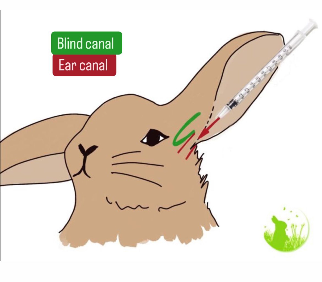
Additional treatment for ear infections in rabbits
In some rabbits, oral mucolytic medication may be considered to help loosen thick secretions. Drugs such as acetylcysteine (NAC) or bromhexine may be used under veterinary supervision. Acetylcysteine may also help reduce the viscosity of thick secretions and can have effects on bacterial biofilms, which may support antimicrobial treatment in selected cases.
Effective pain relief is essential. Meloxicam is commonly used in rabbits and, depending on the severity of the pain, the veterinarian may combine it with additional analgesics such as metamizole or tramadol. The choice, dose and frequency of pain medication must be adjusted to the individual rabbit and its clinical condition.
Systemic antibiotics may also be required for bacterial ear infections, particularly when the infection is severe, difficult to control, or involves the middle ear or deeper tissues.
Good general health, adequate nutrition and appropriate supportive care are important during recovery. A rabbit that is eating less because of pain or illness may require additional nutritional support. Herbal foods such as ginger or horseradish should not be regarded as substitutes for appropriate antimicrobial, anti-inflammatory or analgesic treatment.
Monitoring treatment success
Treatment should be monitored with repeat examinations and, where appropriate, cytology. Medication should not simply be stopped as soon as the rabbit appears better. The veterinarian should confirm that the inflammation and infection have adequately resolved before treatment is discontinued.
This is particularly important because incompletely treated otitis externa can become chronic or progress to otitis media.
Long-term monitoring in lop-eared rabbits
In lop-eared rabbits, recurrent ear disease is often associated with the anatomy of the ear canal. Their narrow, bent ear canals can continue to impair ventilation and normal earwax clearance even after an infection has been successfully treated.
Some affected rabbits therefore require long-term or lifelong ear care, with the frequency of cleaning determined individually by the veterinarian. Regular veterinary ear examinations, including cytology when indicated, can help detect recurrent inflammation at an early stage.
Advanced imaging such as CT, cone-beam CT (CBCT) or MRI may be recommended when middle or inner ear disease is suspected. The need for repeat imaging should be based on the individual rabbit rather than following the same fixed schedule for every animal.
Surgery for recurrent ear infections in rabbits
For rabbits with recurrent or severe otitis externa, particularly lop-eared rabbits with marked narrowing of the ear canal, surgery may be considered. The most appropriate procedure depends on the location and severity of the disease.
- Lateral ear canal resection (Zepp procedure): Part of the lateral wall of the external ear canal is removed to improve ventilation, drainage and access for cleaning or medication. This procedure may be considered for recurrent otitis externa when the disease is primarily confined to the external ear canal.
- Partial ear canal resection: A smaller section of the ear canal wall may be removed in selected cases where the disease is localised.
- Total ear canal ablation: In severe, irreversible disease, the entire external ear canal may need to be removed. This is a considerably more extensive procedure and is generally reserved for advanced cases in which less invasive treatment is unlikely to succeed.
The choice of surgery should be made by a veterinarian or veterinary surgeon experienced in rabbit ear disease, ideally after appropriate diagnostic imaging has established how far the disease extends.
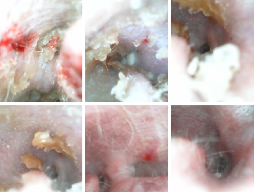
Treatment course of otitis externa in a lop-eared rabbit
Treatment of middle ear infections (otitis media) in rabbits
There are two main approaches to treating otitis media in rabbits:
- Conservative treatment (without surgery): Complete resolution cannot always be achieved. In some cases, treatment suppresses the infection and inflammation, improves the rabbit’s quality of life and keeps the disease under control rather than eliminating it completely. Treatment often needs to continue for several months.
- An effective antibiotic is essential. If the eardrum is damaged or perforated, a sample should ideally be collected from the affected area for bacterial culture and antibiotic susceptibility testing. Material from deeper within the ear may need to be flushed out and submitted for analysis. Superficial samples alone may not accurately reflect the organisms involved. Injectable penicillin may be effective against susceptible bacteria in rabbits, but penicillin must only be used in formulations and by routes that are safe for rabbits. Oral penicillins should not be given to rabbits. Depending on the infection, the veterinarian may select or combine antibiotics with good penetration into deeper tissues. Combinations such as metronidazole and enrofloxacin may be considered in selected cases according to culture results and the individual rabbit.
- Pain management is essential. Meloxicam is commonly used in rabbits and may be combined with additional analgesics such as metamizole or tramadol, depending on the severity of the pain and the rabbit’s clinical condition. Rabbits may show surprisingly few outward signs of pain even with severe middle ear disease, so the absence of obvious pain behaviour does not mean that the condition is painless.
- N-acetylcysteine (NAC) may be used as an adjunct in selected cases. It can help reduce the viscosity of thick secretions and may help disrupt bacterial biofilms, potentially improving drainage and supporting antimicrobial treatment. Depending on the individual case, the veterinarian may use an NAC-containing flushing solution or a Tris-NAC preparation before or alongside topical antimicrobial treatment. Oral mucolytic treatment may also be considered.
- If the rabbit also has otitis externa or the eardrum is not intact, appropriate ear cleaning and flushing are an important part of treatment. The products and technique must be chosen carefully because some substances may be ototoxic if they reach the middle or inner ear.
- Surgical treatment: Recurrence can occur with medical treatment alone, particularly in chronic or advanced cases. Rabbit pus is also extremely thick, so even when the active infection improves, the tympanic bulla may remain filled with material. For this reason, surgery may be recommended in some cases of chronic or severe otitis media. The surgical procedure depends on the location and extent of the disease:
- Ear canal widening surgery in lop-eared rabbits: If chronic external ear disease is contributing to the middle ear infection, widening the external ear canal can improve ventilation and drainage and address the underlying anatomical problem. Intensive flushing and medical treatment of the middle ear may still be required afterwards.
- Myringotomy and drainage: If the eardrum is still intact, a small opening can be made in it (myringotomy) so that material can be suctioned from and the middle ear can be flushed. Follow-up treatment may include appropriate topical antimicrobial medication and repeat imaging, such as CT. This approach is generally most suitable when the bulla has not yet undergone severe thickening or destructive bony changes.
- Lateral bulla osteotomy: This may be considered in advanced disease, particularly when the eardrum is damaged, when otitis media occurs together with otitis externa, or when deeper ear structures are involved. The tympanic bulla is surgically opened, abnormal material is removed, and the cavity is thoroughly flushed. Depending on the surgical technique and individual case, drainage and postoperative flushing may be required while the area heals.
- Ventral bulla osteotomy: This is used less commonly and may be considered when the eardrum is intact and otitis media is present without significant external ear disease. The middle ear is approached surgically from below, allowing the bulla to be opened, cleaned and flushed.
The choice between conservative and surgical treatment should be based on CT or other appropriate diagnostic imaging, the condition of the eardrum and tympanic bulla, the presence of external or inner ear disease, bacterial culture results and the rabbit’s overall condition.
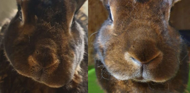
On the follow-up CT scan, the middle ear was still filled with material, but there were no longer signs of active inflammation. Ernie did not experience another relapse, and the ear disease remained clinically inactive for the rest of his life.
This was a case of otitis media without concurrent otitis externa, consistent with an ascending infection from the upper respiratory tract.
Inner ear infection (otitis interna) in rabbits
Otitis interna usually develops as an extension of otitis media and therefore commonly occurs together with a middle ear infection.
- In many cases, intensive treatment of the middle ear infection with antibiotics, mucolytics, betahistine, pain relief and other supportive medication may also improve the inflammation of the inner ear. See the section on otitis media above.
- Severe inner ear infections with neurological signs can be extremely difficult to treat and may not be suitable for surgery. If the rabbit cannot be stabilised and its quality of life is severely compromised, euthanasia may have to be considered. However, some rabbits can stabilise with intensive conservative treatment over several months. Once the condition is stable, surgery may sometimes become an option.
Surgery for ear infections in rabbits – yes or no?
Whether surgery is technically possible and appropriate for an individual rabbit depends on the findings from diagnostic imaging such as CT, cone-beam CT (CBCT) or MRI.
There is currently no single treatment approach that is appropriate for every case. Some veterinarians favour early surgical intervention in an attempt to slow or prevent further progression of the disease. Others recommend surgery only when more advanced changes are present, such as:
- increased pressure within the middle ear or bulging of the eardrum
- thickening, damage or destruction of the tympanic bulla
- clear clinical symptoms
If the bulla is filled with material but the rabbit remains clinically stable and there are no signs of progressive disease, some veterinarians may initially recommend monitoring and conservative treatment.
Surgical options for rabbit ear infections
For lop-eared rabbits with otitis externa only, a comparatively less invasive procedure such as a lateral ear canal resection (Zepp procedure) may be performed. This widens the external ear canal, improves ventilation and drainage, and addresses one of the anatomical factors that predispose lop-eared rabbits to chronic ear disease.
Treating severe external ear disease before it progresses to the middle ear may, in selected cases, reduce the need for more invasive surgery later.
Surgery involving the middle ear, such as a bulla osteotomy, is considerably more invasive and is associated with greater postoperative pain and higher surgical risk. The potential benefits and risks therefore need to be assessed carefully for each individual rabbit.
Prognosis for ear infections in rabbits
Surgery can often bring the inflammatory process under control. Procedures performed for otitis externa generally have a good prognosis, particularly when they correct poor ventilation and drainage of the external ear canal.
The prognosis after surgery for otitis media is more variable. In many rabbits, surgery can lead to resolution of the active inflammation and a substantial improvement in quality of life. However, recurrence is possible, and in some cases surgery may control the disease for a period of time rather than provide a permanent cure.
Some rabbits regain an excellent quality of life after surgery. Rabbits that had previously seemed unusually quiet or inactive because of chronic ear disease may become noticeably more alert and active once pain and inflammation are brought under control.
The prognosis may be somewhat better when the infection has spread from the external ear into the middle ear, as can occur in lop-eared rabbits, than when it has spread from the nasal cavity to the middle ear via the auditory tube, for example in rabbits with chronic respiratory disease.
Neurological signs that are already present before surgery—such as circling, head tilt or facial nerve spasm—may not resolve as a direct result of surgery. However, head tilt can improve considerably in some rabbits with intensive conservative treatment, particularly once the underlying ear disease has been successfully controlled.
Rabbit ear surgery: choosing an experienced surgeon
The experience of the surgeon is one of the most important factors in the success of rabbit ear surgery. These procedures should ideally be performed by a veterinary surgeon with extensive experience operating on rabbit ears. An experienced surgeon is also better able to assess whether surgery is appropriate for the individual rabbit and which procedure offers the best balance of potential benefits and risks.
Rabbit ear surgery is highly specialised, so suitable surgeons can be difficult to find. A rabbit-experienced veterinarian should also coordinate the medical treatment before and after surgery.
For major ear surgery, rabbits should ideally undergo endotracheal intubation during general anaesthesia, and effective pain management is essential.
Aftercare following middle ear surgery
Middle ear surgery is a major procedure and usually requires intensive nursing care for around one to two weeks.
Owners should be prepared to:
- give medication several times a day
- provide assisted feeding if the rabbit is not eating adequately
- attend follow-up veterinary appointments
- monitor the surgical wound closely
- administer eye drops several times a day if eyelid closure is impaired
Good pain management is particularly important. Rabbits in significant pain may stop eating, which can quickly lead to additional gastrointestinal problems.
Possible complications of rabbit ear surgery
Possible complications include:
- wound-healing problems around the ear – these can occur but are often manageable with appropriate treatment
- temporary facial nerve dysfunction, which may affect eyelid closure or cause facial asymmetry or facial nerve spasm
- significant postoperative pain
- reduced appetite or refusal to eat
- head tilt, although this is less common
Facial nerve problems are often temporary and may improve over the following weeks, although recovery varies between individual rabbits.
Lateral ear canal resection / Zepp procedure / “lop-ear surgery”
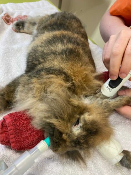
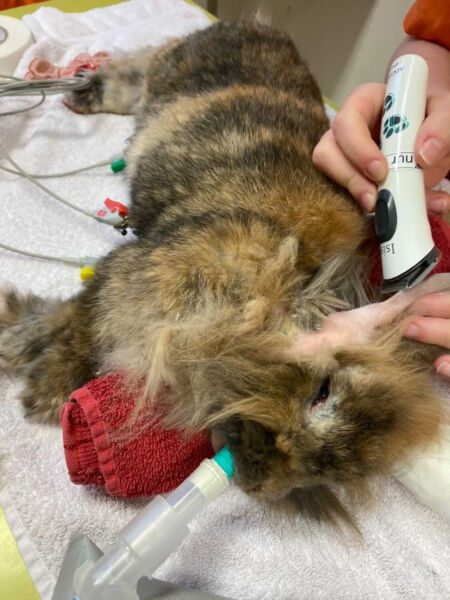
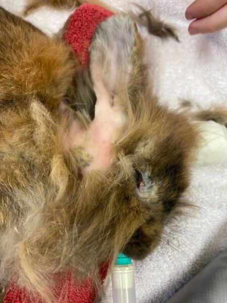
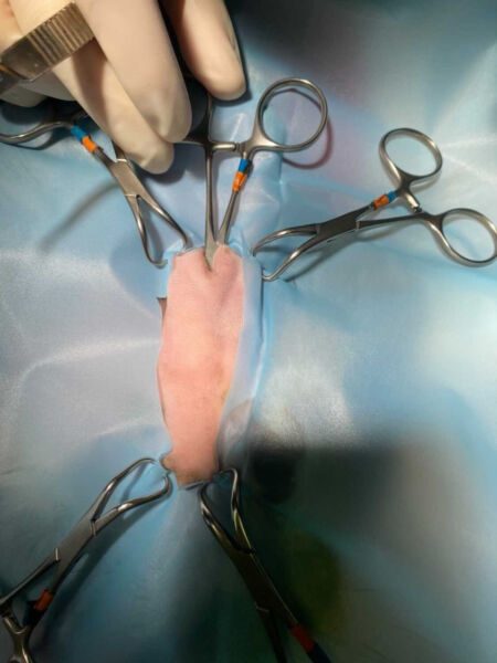
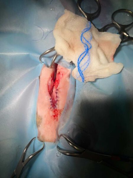
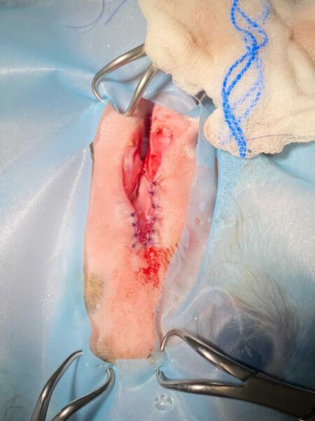
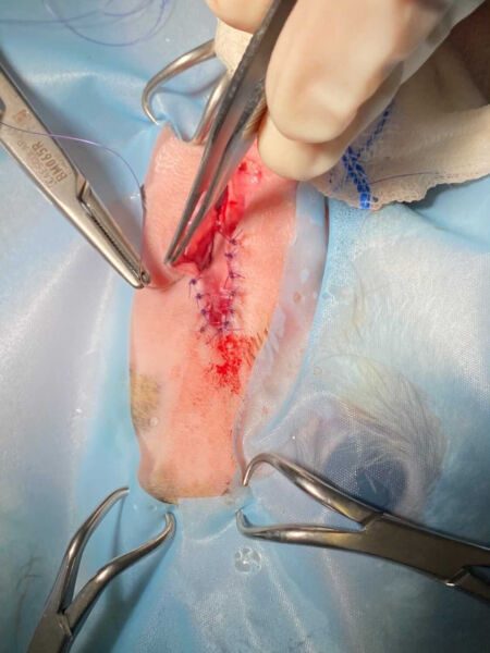
The photos show the rabbit on the day of surgery, at suture removal, and at the follow-up examination two weeks later. The procedure was performed bilaterally, on both ears.
Photos courtesy of Tierarztpraxis Holt / Alexandra Riecken.
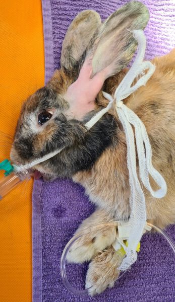
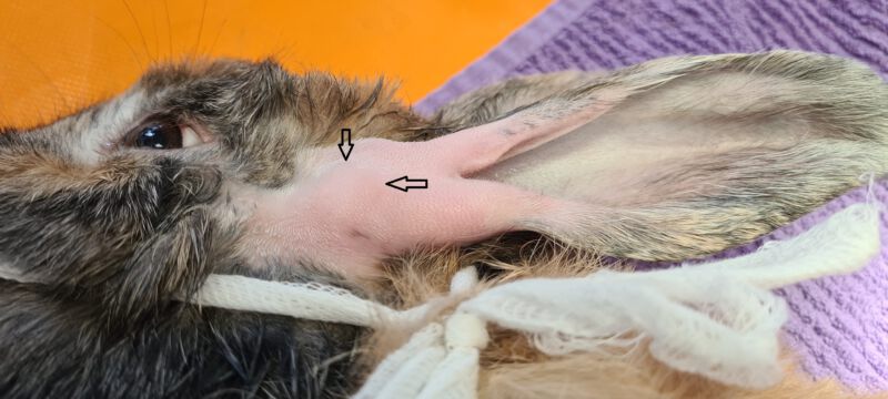
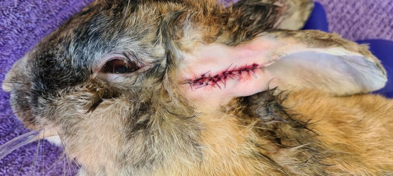
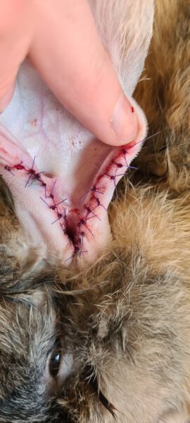
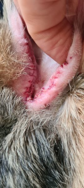
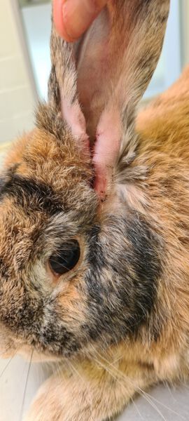
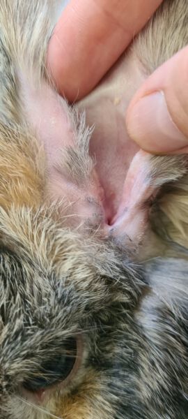
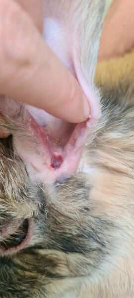
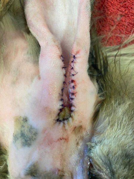
Feivel also underwent surgery for otitis externa. The opening of the external ear canal was widened sufficiently to allow air to reach the area beyond the characteristic bend found in lop-eared rabbits, improving ventilation and allowing the inflammation to heal.
Photos courtesy of Saskia Hinze / Tierarztpraxis Schneidersgarten.
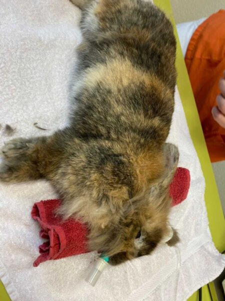
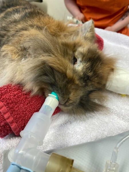
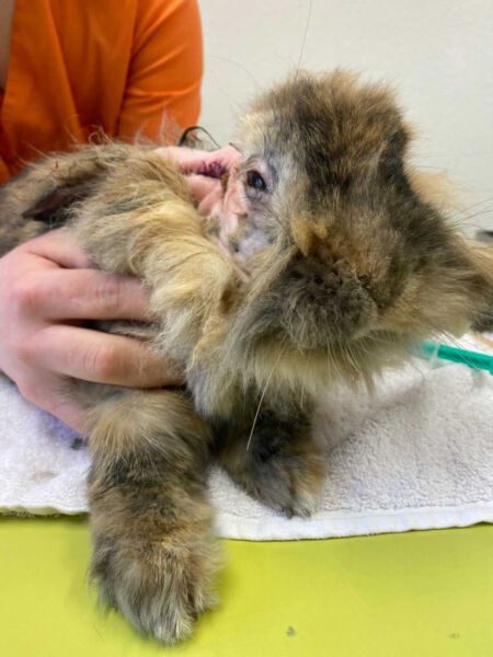
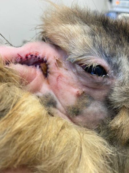
And here is another example of the healing process:
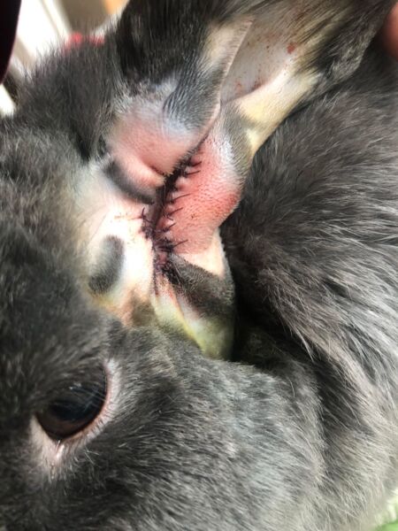
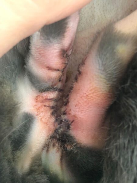
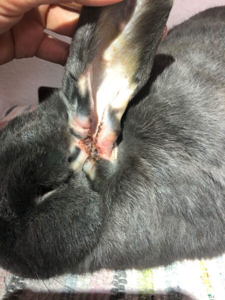
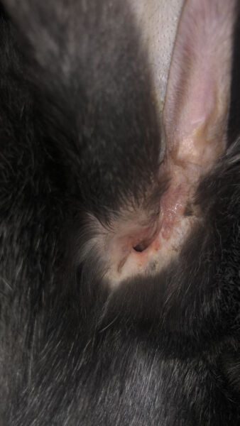
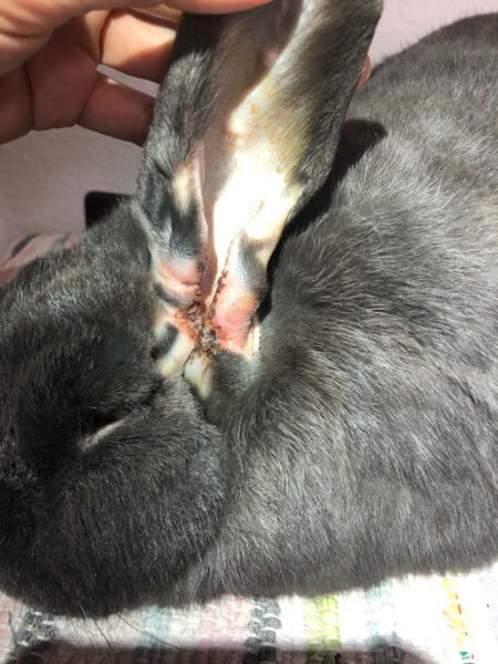
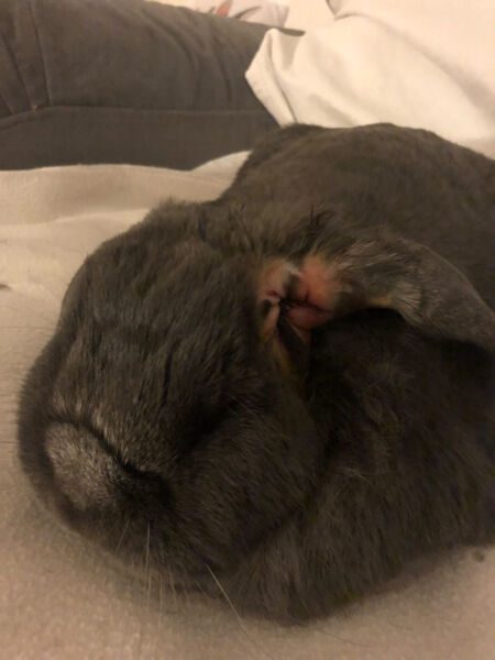
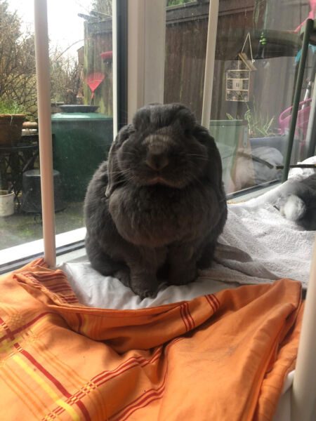
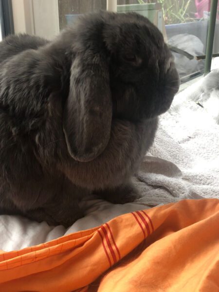
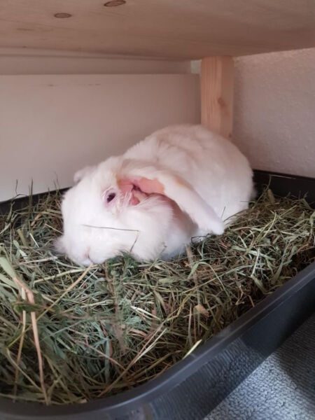
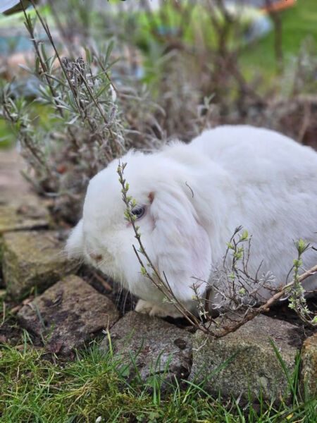
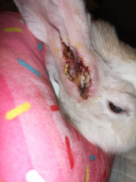
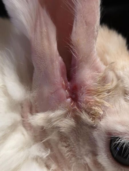
Wound complications are common but manageable. There are 12 days between the first and the last photo:
How to prevent ear infections in rabbits
Lop-eared rabbits are predisposed to both otitis externa and otitis media. CT studies have shown that their ear anatomy increases the risk of chronic ear disease. The hanging pinna, together with the characteristic narrowing and bend of the ear canal, reduces ventilation and interferes with the normal removal of earwax. This creates conditions in which bacteria and yeasts can multiply and contribute to inflammation.
Because these anatomical changes are directly associated with the lop-eared conformation, breeding for pronounced lop ears raises important animal-welfare concerns. Rabbits with chronic respiratory disease or other persistent inflammatory conditions may also be at increased risk of developing otitis.
Rabbits at increased risk may benefit from an individual ear-care and monitoring programme developed with a veterinarian experienced in rabbit medicine.
Ear care for lop-eared rabbits
In some lop-eared rabbits, the underlying anatomical problem remains throughout life. Regular ear care cannot guarantee that inflammation will never develop, but it may help reduce the risk of recurrent or severe disease.
Depending on the condition of the individual ear, preventive care may include:
- Regular ear cleaning, for example weekly or monthly when recommended by the veterinarian. The cleanser should be appropriate for the amount of earwax, the microorganisms present and the condition of the eardrum. Products containing Tris-EDTA may be suitable in selected cases, while ingredients such as chlorhexidine should only be used when appropriate and when the eardrum is known to be intact.
- Thorough cleaning and flushing when medically necessary, sometimes under sedation or during another anaesthetic procedure if the ear canal is severely narrowed or obstructed.
- Monitoring how the rabbit responds to the cleanser. Some preparations can irritate rabbit ears, and even otherwise suitable products may cause redness, discomfort or skin reactions in sensitive animals. If this occurs, the product should be stopped and the veterinarian contacted.
How to detect ear infections early
Early detection is particularly important in lop-eared rabbits, because significant disease may develop before obvious symptoms appear.
Useful monitoring measures include:
- Checking the base of each ear regularly for swelling, warmth, tenderness, itching or differences between the two sides.
- Regular veterinary ear examinations, with cytology when indicated, to detect inflammation or bacterial or yeast overgrowth at an early stage.
- Diagnostic imaging when clinically indicated. CT or cone-beam CT (CBCT) can detect middle ear disease that may not be visible during otoscopic examination or on standard X-rays. MRI may be useful in selected cases, particularly when inner ear, neurological or soft-tissue disease is suspected. The need and frequency of imaging should be determined individually rather than performed on a fixed schedule for every rabbit.
- Knowing the warning signs of ear disease, including head shaking, scratching at the ears, facial asymmetry, head tilt, abnormal head movements, balance problems or nystagmus.
Because rabbits often hide pain and early ear disease can be difficult to detect from the outside, regular monitoring is especially important for rabbits with lop ears or a history of ear problems.
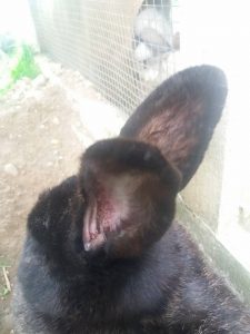
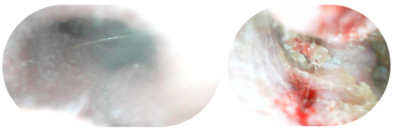
Comparison of a Healthy, Open Ear Canal and an Inflamed, Narrowed Ear Canal
Further Examples:
Mickey with Circular Movements, Snuffles, and Head Tilt
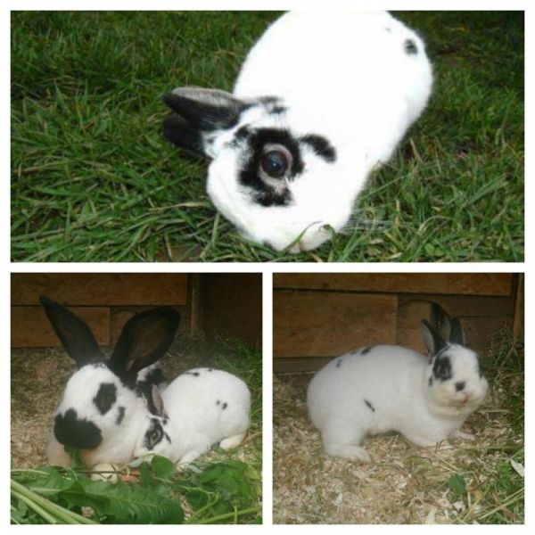
Mickey had a tilted head for 10 months and was initially treated for E. cuniculi (EC) because his blood test showed a positive titer. When his condition didn’t improve, he was left untreated. I decided to pursue detailed diagnostics to determine the cause of his head tilt. It turned out that Mickey had an ear infection. I treated him for both E. cuniculi and the ear infection, and within just a few days, his head straightened completely. His snuffles also improved significantly.
Rabbits with ear infections often tilt their heads to alleviate the severe pain they experience. This is frequently mistaken for E. cuniculi, and proper diagnostics are often overlooked. As a result, many rabbits suffer for months from agonizing pain (anyone who has ever had a severe middle ear infection can imagine how unbearable it must feel).
In Mickey’s case, his ear appeared completely normal from the outside. Even using a camera to inspect the ear canal didn’t reveal anything unusual. The inflammation was only visible on a head X-ray. Blood tests can also indicate signs of inflammation.
Years ago, E. cuniculi was relatively unknown, and it was difficult to find a veterinarian who could diagnose and treat it correctly. Today, however, many issues are mistakenly labeled as “E. cuniculi,” leading to countless animals being incorrectly treated for this condition—even when they are actually suffering from a different, painful disease. As a result, people often say, “My rabbit died of EC.” In reality, rabbits don’t die from E. cuniculi if their kidney function is monitored via blood tests and they receive proper treatment from the beginning. Rabbits can die from kidney failure caused by E. cuniculi, but only if it goes untreated or is mismanaged.
Unfortunately, many rabbits endure immense suffering or die from other diseases that are misdiagnosed as EC.
Mickey was fortunate because his ear infection wasn’t too advanced, and I ensured he received proper diagnostics and treatment. In rabbits with untreated ear infections that persist for months, the infection often leads to bone erosion. In such cases, euthanasia is often necessary, or the animal dies from complications.
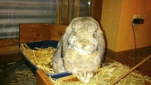
Paul with Crooked Mouth, Head Tilt, and Hind Limb Paralysis
Since July 2015, Paul had a “crooked mouth.” Our veterinarian couldn’t identify the cause or offer a solution, and we couldn’t find any information online either.
Then, in the spring and again in November 2016, Paul’s hind legs suddenly gave way. He was diagnosed with a suspected back problem. As his symptoms didn’t improve, we pushed for further treatment. He was treated twice for E. cuniculi (EC), and both times the symptoms disappeared, and Paul seemed to return to his normal self. However, in November, he developed a slight head tilt that did not fully resolve.
On December 27, 2016, he collapsed again. The following day, we obtained the necessary medications to address what we suspected was an EC flare-up. These included Baytril, Panacur, and a Vitamin B complex. Unfortunately, Paul’s condition worsened significantly, and due to the holiday season, we had to consult a substitute veterinarian.
Paul was examined inadequately, only through palpation and visual inspection. The diagnosis was simply “It must be EC.” He received injections of corticosteroids and enrofloxacin. The treatment had an immediate positive effect, and the next day, he was treated identically. While he improved noticeably, by the weekend, he collapsed again, and we took him to our regular veterinarian the following Monday.
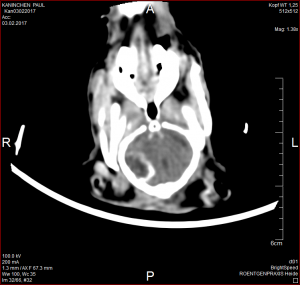
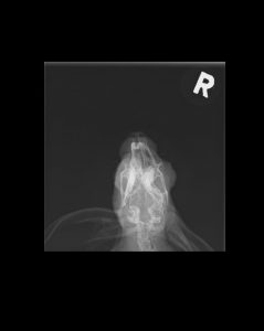
Our regular vet also diagnosed EC and, for the first time, suggested euthanasia, as Paul was barely able to sit up. We insisted on further diagnostics, and a week later, an X-ray was taken and blood tests performed to rule out a middle ear infection (otitis media). Although the X-ray was taken at an awkward angle, it later became clear that it revealed a deformity, which our veterinarian did not recognize. The blood test showed no EC titer and no signs of inflammation.
At this point, Paul needed intensive care for three weeks, as he could only lie down. After our insistence, Paul was treated with Chloramphenicol Palmitate, and we had to hand-feed him. After 10 days, the antibiotic was stopped, and Paul began eating on his own again. He was much improved but could still only lie down.
We sought a second opinion. The new veterinarian performed a CT scan, which revealed a long-standing otitis media and a brain abscess with bone erosion. This was also the cause of his “crooked mouth” (ipsilateral hemifacial spasm/facial nerve paralysis) that had persisted since 2015. The antibiotic treatment, though effective, came about 1¾ years too late.
Sadly, Paul had to be euthanized.
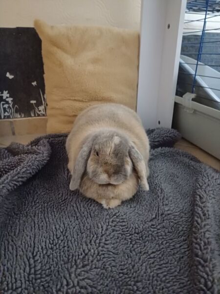
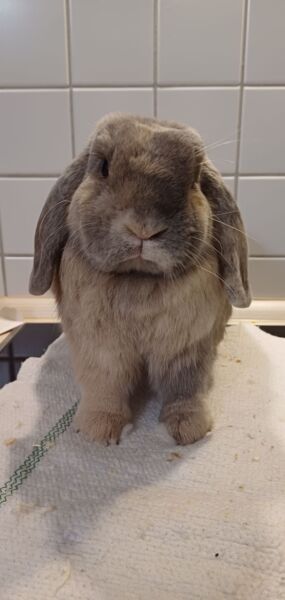
Before-and-After Photos of Left Ear Infection and EC
Frequently asked questions about ear infections in rabbits
Can rabbits have an ear infection without showing symptoms?
Yes. Ear infections in rabbits can remain unnoticed for a long time, because rabbits often show very few outward signs of pain. Some rabbits simply become quieter or less active, while others may show no obvious symptoms at all.
Are lop-eared rabbits more prone to ear infections?
Yes. Lop-eared rabbits have an increased risk of ear disease because their ear canals are typically narrower and bent. This can reduce ventilation and interfere with the normal removal of earwax, creating conditions that favour chronic inflammation.
Can an ear infection cause head tilt in rabbits?
Yes. Middle and inner ear infections can cause head tilt, balance problems, circling and nystagmus when structures involved in balance are affected.
Can an ear infection be mistaken for E. cuniculi?
Yes. A rabbit with neurological signs may have E. cuniculi, an ear infection, or both conditions at the same time. The symptoms can overlap considerably, particularly head tilt, balance problems, circling and abnormal eye movements.
Because of this overlap, some rabbits are diagnosed with presumed E. cuniculi without the ears being investigated thoroughly enough. A positive E. cuniculi antibody titre does not by itself prove that E. cuniculi is responsible for the current symptoms.
For rabbits with neurological signs, middle and inner ear disease should therefore always be considered and appropriately investigated, often with diagnostic imaging such as CT or MRI.
How are ear infections diagnosed in rabbits?
The external ear canal can be examined with an otoscope or endoscope, but in Mini Lops and other lop-eared rabbits the characteristic bend and narrowing of the ear canal can make the deeper part of the canal difficult or impossible to see. As a result, the visible part of the ear may appear clean even though inflammation, excessive earwax or infection is present beyond the bend.
For this reason, CT is particularly valuable in lop-eared rabbits, because it allows the veterinarian to assess the entire ear, including the deeper external ear canal, middle ear and surrounding structures. It can show whether the ear canals are clear, how far earwax or inflammatory material extends, and whether one or both middle ears are affected.
In rabbits with recurrent ear problems or a high risk of chronic ear disease, regular imaging may be recommended. Whether CT should be performed annually should be decided individually with a rabbit-experienced veterinarian rather than using the same schedule for every rabbit.
Can ear infections in rabbits be treated without surgery?
Sometimes. Conservative treatment with appropriate medication, pain relief, ear cleaning and flushing may successfully control some ear infections. Chronic, recurrent or advanced disease may require surgery.
How long does treatment for an ear infection in rabbits take?
Treatment often lasts several weeks, while chronic or complicated middle ear infections may require treatment for several months. The duration depends on the location and severity of the infection and how well the rabbit responds to treatment.
Can ear infections come back?
Yes. Recurrence is particularly common in rabbits with chronically narrow or poorly ventilated ear canals, especially lop-eared rabbits. Some rabbits therefore require long-term monitoring and regular ear care.
About This Article
Original content by Viola Schillinger, veterinarian
Veterinarian specializing in rabbit health, nutrition and species-appropriate husbandry, and founder of Kaninchenwiese.de, a long-established German rabbit care and health resource reaching around 500,000 visitors per month.English translation and adaptation by Erika Kühne Escobar
Medically reviewed by Viola Schillinger, veterinarian
Last medically reviewed: August 2026
Sources, including:
Anonymous (2018): Qualzucht beim Kaninchen – Leiden für die Schönheit? Kugelköpfe, Schlappohren und Lebensschwäche, VetImpulse Nr. 13, 27. Jg.
Capello, V. (2006): Lateral ear canal resection and ablation in pet rabbits. In: North American Veterinary Conference 20, 2006, Orlando, Florida, S. 1711-1713.
Capello, V. (2018). Ear surgery of pet rabbits. In BSAVA Congress Proceedings 2018 (pp. 50-51). BSAVA Library.
Chivers, B. D., Keeler, M. R., & Burn, C. C. (2023). Ear health and quality of life in pet rabbits of differing ear conformations: A UK survey of owner-reported signalment risk factors and effects on rabbit welfare and behaviour. PloS one, 18(7), e0285372.
Chow, E. P. (2011): Surgical management of rabbit ear disease. Journal of Exotic Pet Medicine, 20(3), 182-187
Chow, E. P., Bennett, R. A., & Dustin, L. (2009): Ventral bulla osteotomy for treatment of otitis media in a rabbit. Journal of Exotic pet medicine, 18(4), 299-305.
Csomos, R., Bosscher, G., Mans, C., & Hardie, R. (2016): Surgical management of ear diseases in rabbits. Veterinary Clinics: Exotic Animal Practice, 19(1), 189-204.
Díaz, L., Castellá, G., Bragulat, M. R., Martorell, J., Paytuví-Gallart, A., Sanseverino, W., & Cabañes, F. J. (2021). External ear canal mycobiome of some rabbit breeds. Medical Mycology, 59(7), 683-693.
Dinicola, S., De Grazia, S., Carlomagno, G., & Pintucci, J. P. (2014): N-acetylcysteine as powerful molecule to destroy bacterial biofilms. A systematic review. Eur Rev Med Pharmacol Sci, 18(19), 2942-2948.
Dyckhoff, G., Hoppe-Tichy, T., Kappe, R., & Dietz, A. (2000): Antimykotische Therapie bei Otomykose mit Trommelfelldefekt. HNO, 48(1), 18-21.
Cole LK, Papich MG, Kwochka KW et al. (2009): Plasma and ear tissue concentrations of enrofloxacin and its metabolite ciprofloxacin in dogs with chronic end-stage otitis externa after intravenous administration of enrofloxacin. Vet Dermatol 2009; 20: 51–59
Eatwell, K. (2013): Diagnosis of otitis externa, media and interna in rabbits. Veterinary Times, 43(13), 20-22.
Eatwell, K, Mancinelli, E., Hedley, J., Yool, J. (2013): Partial ear canal ablation and lateral bulla osteotomy in rabbits
Eckert, Y., Witt, S., Reuschel, M., & Fehr, M. (2017): Otitis beim Kaninchen–Symptome, Diagnostik und Therapiemöglichkeiten. kleintier konkret, 20(S 02), 2-9.
Ewringmann, A. (2016): Leitsymptome beim Kaninchen: Diagnostischer Leitfaden und Therapie. Georg Thieme Verlag
Farca, A. M., Piromalli, G., Maffei, F., & Re, G. (1997): Potentiating effect of EDTA‐Tris on the activity of antibiotics against resistant bacteria associated with otitis, dermatitis and cystitis. Journal of small animal practice, 38(6), 243-245.
Flatt, R. E.; Deyoung, D. W.; Hogle, R. M. (1977): Suppurative otitis media in the rabbit: prevalence, pathology, and microbiology. Laboratory animal science, 27. Jg., Nr. 3, S. 343-347.
Galuppi, R., Morandi, B., Agostini, S., Dalla Torre, S., & Caffara, M. (2020): Survey on the presence of Malassezia spp. in healthy rabbit ear canals. Pathogens, 9(9), 696.
Haberfield, J. (2015): Otitis Media in Rabbits.
Johnson, J. C., & Burn, C. C. (2019): Lop-eared rabbits have more aural and dental problems than erect-eared rabbits: a rescue population study. BioRxiv, 671859.
Liebscher, J., & Hein, J. (2023): Typische und untypische Infektionserreger beim Kaninchen: Teil 5 Neurologische Symptome. kleintier konkret, 26(S 01), 12-17.
Mäkitaipale, J., Harcourt-Brown, F. M., & Laitinen-Vapaavuori, O. (2015): Health survey of 167 pet rabbits (Oryctolagus cuniculus) in Finland. Veterinary Record, vetrec-2015.
Mancinelli, E., Lennox, A. M. (2017): Management of Otitis in Rabbits. Journal of Exotic Pet Medicine 26 (2017), pp 63–73
Maruhashi E, Braz BS, Nunes T, Pomba C, Belas A, Duarte-Correia JH, Lourenço AM (2016):Efficacy of medical
grade honey in the management of canine otitis externa – a pilot study. Vet Dermatol. 27(2):93-8e27.
Meredith, A., & Lord, B. (2014). BSAVA manual of rabbit medicine. British Small Animal Veterinary Association
Matos, R., Ruby, J., Hatten, R. A. van, Thompson, M. (2015): Computed tomographic features of clinical and subclinical middle ear disease in domestic rabbits (Oryctolagus cuniculus): 88 cases (2007–2014). Journal of the American Veterinary Medical Association, February 1, 2015, Vol. 246, No. 3 , Pages 336-343 [https://doi.org/10.2460/javma.246.3.336, 27.08.2017]
Mayer, J. (2011): Otology of the rabbit: Anatomy, Physiology and Surgery. Saturday Proceedings of the Association of Exotic Mammal Veterinarians congress, Seattle, USA. pp47-52
Reuschel, M. (2017): Ohrentzündungen bei Kaninchen. [https://vetline.de/ohrentzuendungen-bei-kaninchen/150/3252/103523/, 15.09.2017]
Reuschel, M. (2018): Untersuchungen zur Bildgebung des Kaninchenohres mit besonderer Berücksichtigung der Diagnostik einer Otitis bei unterschiedlichen Kaninchenrassen.
Richardson, J., Longo, M., Liuti, T., & Eatwell, K. (2019): Computed tomographic grading of middle ear disease in domestic rabbits (Oryctolagus cuniculi). Veterinary Record, vetrec-2018.
Ruby, J., Van, R. H., & Thompson, M. (2015): Computed tomographic features of clinical and subclinical middle ear disease in domestic rabbits (Oryctolagus cuniculus): 88 cases (2007-2014). Journal of the American Veterinary Medical Association, 246(3), 336-343.
Thöle M, Parmentier S, Müller LS (2020): Ohrerkrankungen beim Heimkaninchen (Oryctolagus cuniculus) – Teil 1: Anatomische Grundlagen und diagnostische Aufarbeitung. Kleintierprax 65 (09): 489–506
Varga, M. (2014): Textbook of Rabbit Medicine. Second Edition